A recent research has found that coarse grained small hydrophobic graphene sheets pierce through the phospholipid membrane and can wreak havoc with the membrane of bacteria. Once the bacteria membrane has been breached, the bacteria have difficulty functioning. While the material has the ability to thwart the growth of some bacterial strains, cells in mammals are not harmed. The research team specifically used the material to test different species of bacteria associated with tooth decay and gum disease. They concluded that graphene oxide limited the growth of pathogens by destroying the bacterial walls and membranes. For this reason, the researchers thought this material would be useful in dentistry.
Archives for March 2015
T-cleanA can help with your oral hygiene
The positive effects of hydrogen peroxide on oral inflammation has been known for many years. The challenge with 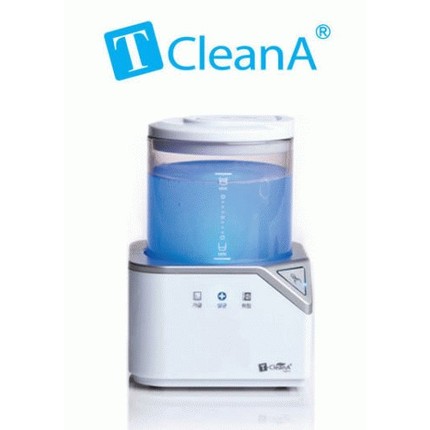 using hydrogen peroxide is that the active ions do not last for long. We have now available for sale a special bath which can generate hydrogen peroxide from tap water at your home, where it can be put to the following uses:
1.For cleaning and disinfecting sport mouthguards, nightguards and orthodontic appliances.
2. For rinsing in conjunction with brushing and flossing to reduce gingival inflammation.
3. For cleaning dentures like denture tablets.
For more information click here.
Molar Incisor hypomineralization may result from exposure to BPA
A recent research found that tooth enamel abnormality in children, molar incisor hypomineralization (MIH), may result from exposure to the industrial chemical bisphenol A (BPA). The authors of a new study reached this conclusion after finding similar damage to the dental enamel of rats that received BPA.
These result may shed light on the increasing prevalence of molar and incisor hypomineralization in the developed world. Hopefully with the increase in awareness to the presence of BPA and it’s effects this trend will stop.
For more detailed way of dealing with the consequences of molar and incisor hypomineralization please read this post.
How healthy are your gums?
The condition of the gums has many consequences on your general health. Gum disease can lead to diabetes, stroke, premature babies and other medical conditions on top of the local effects in the oral cavity.
Recently the European Federation of Periodontology has published an easy with list of questions for self assessment of the gum condition. Please press this link to take the test.
Please don’t hesitate to contact us for more information about gum disease or to make an appointment to examine the condition of gums.
Best mouth guard for tooth clenching and grinding?
A night guard, also known as an occlusal splint, is an appliance that protects the teeth and other vital structures by offering a barrier between the upper and lower teeth. In a large review of the published literature it has been found that the occlusal splint is proven to prevent tooth loss. There are two important features of the splint, the material it is made of and it’s design, that impact its protective function.
Since one size does not fit all, the splints bought at the chemist have to be molded. That is the reason, they are made of a soft material. It has been claimed that a guard of a soft material, is better because it can cushion the impact on the teeth. But this concept has not been scientifically proven, Dr. Kaufman’s experience shows that a soft material caves in while a hard material dissipates the forces over a larger area, therefore reducing the impact of clenching and grinding forces to individual teeth. Leading to the conclusion that a hard occlusal splint would be better at tooth protection. The resilient mouth guards will allow for a perfect adaptation between the teeth and the splint, prevent pain from misfitting splints and avoid the loosing the splint easily.
Another property of the chemist splint is that they are thick. A recent study has shown that the soft mouth guard needs to be at least 4 mm thick for it to protect the teeth. This finding led the researchers to the conclusion that the thick mouth guards are uncomfortable to wear . While the hard acrylic splints Dr. Kaufman provided are thin to allow good protection without discomfort.
Since we have little control on clenching and grinding and it can arise from a variety reasons, we need to concentrate on protecting the teeth. A custom made splint of hard material is the ideal solution.
For an appointment to prescribe the right splint for you please contact us.
How to deal with morning bad breath?
As we sleep, our salivary glands are less active, but the bacteria are not. Our saliva rinses the mouth and washes away the bacteria that cause bad breath, it helps the ingestion and breakdown food particles and removes them from the mouth. People who normally don’t breathe through their mouth may do so while they sleep, drying the oral tissues even further. Another reason for bad breath is, the thirst and starvation condition the body is in while we sleep. For these reasons there is a high risk of having an embarrassing bad breath when we wake up in the morning.
To prevent the bad breath we need to deal with the underlying reasons by:
- Brush and floss our teeth before we go to bed. At time Dr. Kaufman will prescribe other appliances or products to achieve a clean mouth.
- For some individuals, like smokers, there are large deposits of bacteria hiding in the folds of the tongue, that can be removed with a tongue scraper.
- Drink water before you go to bed and have a bottle next to your bed if you feel your mouth is dry at night.
- Limit sugar intake before bedtime, to decrease the available food for the bacteria.
Please don’t hesitate to contact us for more information about bad breath or to schedule an appointment to examine the condition of the dentition .
When to stop thumb sucking?
Many times I’m asked by parents, when do they need to halt the thumb sucking habit of their child?
Babies have natural sucking reflexes, which leads them to put their thumbs or fingers into the mouth,  because it is soothing . Sometimes they develop a habit of thumb sucking when they’re bored, tired or anxious. Thumb sucking becomes a concern when the permanent teeth come into play. At this point the habit might begin to affect the roof of the mouth, the position of the tongue, the way the lip functions or how the teeth are positioned. The point when the parents need to become concerned is when the childs reaches the age of 3.5 years old since it may affect the speech and cause lisping, cause a “trapped lower lip” kind of swallowing, prevent the lower jaw from developing in the right direction and affect the position of the permanent teeth.
To help the child stop the habit it is always good to use positive reinforcements. Give them reminders and possibly set targets that will help the child lose the habit. At times it important to identify the reason for sucking, like feeling more secure and trade the sucking with a cuddly animal.
Please don’t hesitate to contact us for more information about sucking or to schedule an appointment to examine the condition of the dentition .
Read the label for Sugar!
The amount of sugar contained within our most popular breakfast cereals, has been brought to the public’s attention. Analysis by the Obesity Policy Coalition reported in the papers show that four of the 10 most popular children’s cereals contain more than 30% sugar. That means that in a 100g serving there are eight teaspoons of sugar. The guidelines for consumption of added sugars are for no more than 10% for someone who is not overweight. The World Health Organisation recommends less than 5% of sugar, for “additional health benefits”. In other words to minimise the risk of obesity, heart disease and some cancers this is the target that we should be adopt. It demonstrates again how the healthy diet image, we are sold, may be not so healthy and we need to be vigilant and read the ingredients on the label to tell what we are ingesting. We know what sugar does to our teeth and its contribution to the risk for decay but if that energy isn’t used for motion our body converts it to fat and it adds to the risk of obesity and diabetes.
Tooth clenching, Tooth grinding or bruxism
Stress is an unfortunate part of our life, one of its manifestations is tooth clenching and grinding. Our mouth has evolved in such a way that it stays slightly ajar and the teeth don’t touch usually. This way the teeth can retain the strong enamel cap for longer and it is not worn down. The only time teeth are meant to come together is when chewing or swallowing. When we are stressed, among other things our main chewing muscles contract and the teeth meet and press against each other, this is clenching. In some of us in addition to clenching other muscles start to pull the lower jaw forward and sideways leading to the grinding motion or bruxism. This process can be triggered by other lifestyle factors like smoking, large caffeine intake and heavy alcohol consumption or by taking antidepressants.
The clenching leads to the formation of minute cracks in the enamel which over time develop to larger visible cracks and extend to the dentine which is the inner layer of the tooth. The grinding or bruxing of the teeth leads to tooth chipping, wear and loss of tooth. Once the teeth have cracked or have been lost they will not heal back like broken skin or broken bone and they will continue to deteriorate. To prevent the destruction we need to separate the teeth when they are clenched and guide them in non-destructive paths when the muscles pull the jaw sideways. This is achieved with an occlusal splint or a night guard.