Tooronga Family Dentistry in Glen Iris

Family dental care in Glen Iris

Book Now

98227006
0450067475

Suite 1.02, 1 Crescent Rd., Glen Iris 3146
  • About us
    • Dr Daniel Kaufman – Experienced Dental Care Professional in Glen Iris
    • Our Practice in Glen Iris
    • State of the art materials and equipment in Glen Iris
    • 10 reasons to visit us in Glen Iris
    • Strict Infection Control at Tooronga Family Dentistry in Glen Iris
  • Services
    • Orthodontic braces
    • Straight Teeth with Clear aligners in Glen Iris
      • Invisalign Clear Aligners: A Discreet Alternative to Braces in Glen Iris
    • Wisdom tooth extraction
    • Fixing broken teeth.
    • Kids Dentist in Glen Iris
    • Healing bleeding Gums and bad breath
    • Mending teeth with a large cavities.
    • Teeth Whitening
    • Dealing with dental fear
    • Eliminating bad breath – Halitosis
    • Replacing missing teeth
    • Reinforcing fragile teeth in Glen Iris
    • Treating Molar and Incisor Hypomineralization
  • Contact
    • Emergency Contact
    • Book an Appointment Online
    • Ask Dr Kaufman a question
  • Patient information
    • Our Practice in Glen Iris
    • Blog
    • What is a Root canal treatment
    • Why remove decay from teeth
    • Oral hygiene in Glen Iris
    • Gum Disease During Pregnancy in Glen Iris: Causes, Symptoms, and Risks
    • Crowns and Bridges in Glen Iris
    • What to do for traumatic tooth injuries in Glen Iris
    • Tooth removal – Extractions
    • Child Dental Benefits Schedule
    • Professionaly made mouthguard
    • Practice policies in Glen Iris
    • Privacy Policy
    • Terms and Conditions
    • Sitemap

Archives for May 2015

Save Your Tooth from Extraction in Glen Iris: Why “Bad Teeth” Can Almost Always Be Saved

Posted on 05.28.15

Frequently, young patients come to me asking for a tooth to be removed because their teeth are bad. But are they really bad, and is removing the tooth the best way to go?
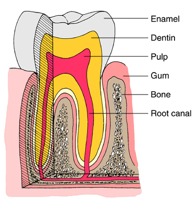
 At Tooronga Family Dentistry, Dr. Kaufman encounters this scenario regularly—Glen Iris patients in their 20s and 30s convinced their damaged teeth are beyond saving, requesting extraction as the “simplest solution.” The reality? The broken-down teeth are usually consequences of two common diseases—decay and gum disease—both caused by bacteria. The crucial insight: their removal will sort things out. Understanding that there are no bad teeth—only teeth damaged by treatable bacterial infections—transforms the conversation from “remove it” to “let’s save it.” Because removing the bacteria and dealing with the causes of tooth breakdown will keep them in your mouth, while extraction creates a cascade of problems far worse than the original damage.

Let’s explore why tooth preservation should almost always be the first choice—and what happens when teeth are unnecessarily removed.


The “Bad Teeth” Misconception

Reframing the problem:


What Patients Mean by “Bad”:

When young Glen Iris patients say their teeth are “bad,” they typically describe:

⚠ Large cavities (visible holes, dark areas) ⚠ Fractured teeth (broken cusps, missing portions) ⚠ Discolored teeth (dark, stained—assuming death/disease) ⚠ Painful teeth (infection, abscess) ⚠ Loose teeth (mobility from gum disease) ⚠ Embarrassing teeth (social stigma—affecting confidence)


The Emotional Response:

Why patients request extraction:

✓ Shame (feeling they’ve “failed” at dental care) ✓ Hopelessness (assuming damage irreversible) ✓ Fear of judgment (anticipating criticism from dentist) ✓ Cost anxiety (believing restoration too expensive—extraction cheaper) ✓ Pain desperation (wanting immediate relief—extraction seems fastest) ✓ Resignation (“Just get rid of it”)

Dr. Kaufman’s perspective:

These teeth aren’t “bad”—they’re damaged by disease. There’s a critical difference:

  • Bad suggests inherent defect (nothing can be done)
  • Diseased indicates treatable condition (intervention can restore health)

The Reality: Treatable Diseases, Not Hopeless Teeth

Understanding the root causes:

“The broken-down teeth are usually consequences of two common diseases.”


Disease #1: Dental Decay (Cavities)

The bacterial assault:

“The first is decay, which is a bacterial assault on our teeth fueled by sugars from our die
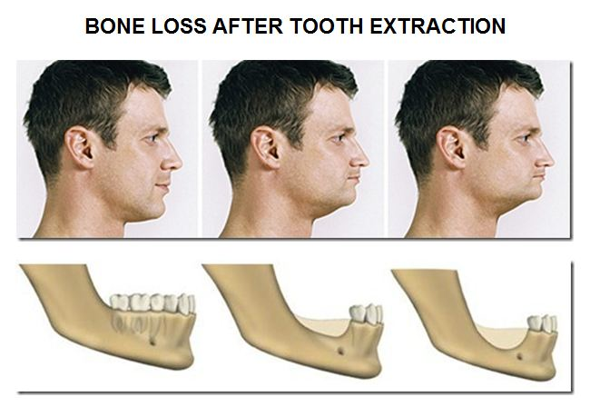
t.”


The Decay Process:

How cavities form:

  1. Bacteria colonize teeth (Streptococcus mutans, others—forming plaque biofilm)
  2. Sugars consumed (from diet—sweets, carbohydrates, sugary drinks)
  3. Bacteria metabolize sugars (producing lactic acid as byproduct)
  4. Acid attacks enamel (dissolving mineral crystals—demineralization)
  5. Cavity forms (hole in tooth structure—progressive enlargement)
  6. Decay advances (through enamel → dentin → pulp if untreated)

Why Young Patients Particularly Affected:

Risk factors common in 20s-30s:

⚠ Dietary habits (frequent snacking, energy drinks, soda consumption) ⚠ Busy lifestyles (irregular brushing, skipping dental visits) ⚠ Limited resources (early career—delaying treatment due to cost) ⚠ Previous childhood decay (multiple fillings—weakened tooth structure vulnerable) ⚠ Genetic susceptibility (some individuals’ enamel/saliva more cavity-prone)

Glen Iris young professionals often present with extensive decay—years of accumulation from adolescence/young adulthood without consistent dental care.


The Key Insight: Decay Is Bacterial

“These problems are caused by bacteria.”

Implications:

✓ Bacteria = treatable (can be removed through dental procedures) ✓ Not structural defect (tooth itself fundamentally sound—just infected) ✓ Reversible damage (removing bacteria, restoring structure saves tooth) ✓ Preventable recurrence (controlling bacteria prevents future decay)

The solution isn’t extraction—it’s bacterial removal + restoration.


Disease #2: Gum Disease (Periodontitis)

The soft tissue infection:

“The second is gum disease caused by bacteria colonizing the soft tissue around the teeth and making an infection there.”


The Gum Disease Process:

Progression:

  1. Bacteria accumulate (plaque below gum line)
  2. Gum inflammation (gingivitis—red, swollen, bleeding gums)
  3. Pocket formation (gums detach from teeth—creating spaces)
  4. Bacterial invasion (deeper colonization in pockets)
  5. Immune response (body attacks bacteria—collateral damage to bone, ligament)
  6. Bone destruction (supporting bone dissolves—tooth loses anchorage)
  7. Tooth mobility (loose teeth—advanced stage)

The Systemic Danger:

“Which can penetrate further to the bone, blood system, and from there to the whole body.”

Beyond the tooth:

⚠ Bone penetration (osteomyelitis—bone infection, rare but serious) ⚠ Bloodstream entry (bacteremia—bacteria circulating systemically) ⚠ Whole body impact (cardiovascular disease, diabetes complications, rheumatoid arthritis, stroke, pregnancy complications)

Glen Iris patients with gum disease experience not just local tooth problems but systemic health threats—making treatment even more critical.


The Key Insight: Gum Disease Is Bacterial

Again—caused by bacteria:

✓ Remove bacteria → Infection resolves ✓ Bone can stabilize (stops destroying further) ✓ Teeth firm up (reduced mobility as inflammation decreases) ✓ Systemic health improves (eliminating bacterial reservoir)

The solution isn’t extraction—it’s periodontal therapy (deep cleaning, antibiotics, ongoing maintenance).


The Solution: Bacterial Removal, Not Tooth Removal

Treating the cause:

“Their removal will sort things out.”

Note: “Their” refers to bacteria removal, not tooth removal—critical distinction.


For Decayed Teeth:

Bacterial removal + restoration:


Step 1: Remove Decay

✓ Excavation (drilling out infected tooth structure) ✓ Disinfection (antimicrobial rinses, medicaments) ✓ Complete removal (all bacteria-laden decayed tissue eliminated)


Step 2: Restore Structure

Depending on extent:

✓ Fillings (composite, amalgam—small to moderate cavities) ✓ Onlays/inlays (larger cavities—partial coverage) ✓ Crowns (extensive decay—full coverage protection) ✓ Root canal + crown (if decay reached pulp—saving tooth from extraction)

Even severely decayed teeth—those patients assume “hopeless”—can often be saved with root canal therapy and crown restoration.


For Gum Disease:

Periodontal therapy:


Step 1: Deep Cleaning

✓ Scaling and root planing (removing bacteria, calculus below gum line) ✓ Pocket irrigation (antimicrobial rinses flushing bacteria) ✓ Antibiotics (topical or systemic—eliminating resistant bacteria)


Step 2: Maintenance

✓ Frequent cleanings (every 3-4 months—preventing recolonization) ✓ Improved home care (proper brushing, flossing technique) ✓ Monitoring (pocket depth measurements tracking improvement)

Even loose teeth from gum disease can firm up with aggressive periodontal treatment—avoiding extraction.


The Paradigm Shift:

“So there are no bad teeth, removing the bacteria and dealing with the causes to tooth breakdown will keep them in your mouth.”

What this means:

✓ Teeth aren’t inherently defective (they’re diseased—treatable) ✓ Bacterial control is key (addressing root cause) ✓ Prevention possible (controlling bacteria prevents recurrence) ✓ Preservation achievable (even extensively damaged teeth salvageable)

Glen Iris patients should understand: your teeth want to survive—given proper treatment (bacterial removal, structural restoration, ongoing maintenance), they will.


The Extraction Cascade: Why Removing Teeth Makes Things Worse

The domino effect:

“Having the teeth removed will only make things worse for the remaining ones.”


Problem #1: Overloading Remaining Teeth

“By overloading them”:


The Biomechanics:

Normal dentition:

✓ 28-32 teeth (distributing chewing forces) ✓ Each tooth handles portion of load ✓ Force per tooth: Manageable (within physiological tolerance)

After extraction:

⚠ Fewer teeth (remaining teeth must compensate) ⚠ Same total force (from jaw muscles—doesn’t decrease) ⚠ Force per tooth increased (concentrated on survivors) ⚠ Overload damage (accelerated wear, fractures, mobility)


The Vicious Cycle:

  1. First tooth extracted (patient believes problem solved)
  2. Remaining teeth overloaded (handling extra force)
  3. Overload causes damage (fractures, increased decay from food trapping, gum disease from cleaning difficulty)
  4. Second tooth fails (requires extraction)
  5. Further overload (distributed among even fewer teeth)
  6. Third tooth fails → Fourth → Fifth…
  7. Cascade continues until extensive tooth loss

Glen Iris patients who extracted “one bad tooth” at 25 often present at 35 with multiple additional failures—each extraction accelerating the next.


Problem #2: Tooth Migration

“Leading them to move”:


Natural Tooth Stability:

Teeth held in position by:

✓ Adjacent tooth contact (neighbors preventing drift) ✓ Opposing tooth contact (upper and lower preventing vertical movement) ✓ Periodontal ligament (anchoring tooth in socket)

Balance maintained when all teeth present.


What Happens After Extraction:

Loss of stability:

⚠ Tooth behind gap drifts forward (tipping into space) ⚠ Tooth in front of gap drifts backward ⚠ Opposing tooth overerupts (grows into space—no contact stopping it)

Timeline:

  • Months 1-6: Tipping begins (subtle—often unnoticed)
  • Months 6-12: Obvious drift (gap closing, opposing tooth elongated)
  • Years 1-5: Severe malposition (teeth significantly displaced)

Consequences of Migration:

⚠ Food trapping (new spaces between shifted teeth—difficult cleaning) ⚠ Gum disease (trapped food → bacterial growth → periodontitis) ⚠ Decay (food impaction areas vulnerable to cavities) ⚠ Bite problems (shifted teeth don’t contact properly—TMJ issues) ⚠ Adjacent tooth damage (overerupted tooth hitting incorrectly—trauma) ⚠ Orthodontic problems (correcting drift requires braces—expensive, time-consuming)


Problem #3: Chewing Difficulty

“And make chewing difficult”:


Functional Impairment:

With missing teeth:

⚠ Reduced chewing efficiency (fewer teeth = less effective grinding) ⚠ Avoidance of certain foods (tough meats, raw vegetables—too difficult) ⚠ Digestive problems (inadequately chewed food → GI issues) ⚠ Nutritional deficiency (limited diet → vitamin/mineral insufficiency) ⚠ Weight changes (difficulty eating—unintended weight loss) ⚠ Social withdrawal (embarrassment eating in public)


Specific Scenarios:

Losing molars:

  • Chewing function most compromised (molars provide 90% of grinding power)
  • Shift to front teeth (not designed for grinding—damage accelerates)

Losing front teeth:

  • Biting function lost (can’t bite sandwiches, apples)
  • Speech affected (lisp, whistling—/s/, /f/, /th/ sounds)
  • Aesthetic devastation (visible gap—social/professional impact)

Glen Iris young professionals missing teeth often report career impacts (avoiding presentations, client meetings—self-consciousness about appearance).


Problem #4: Bone Loss and Facial Changes

“Once the teeth are removed, the bone that surrounds them will go, leading to changes in your appearance that make you look older.”


The Bone Resorption Process:

Why bone disappears:

✓ Teeth stimulate bone (chewing forces transmitted through tooth root → bone responds by maintaining density) ✓ No tooth = no stimulation (bone receives message “not needed”) ✓ Bone resorbs (body reabsorbs bone tissue—use it or lose it)

Timeline:

  • First year: 25% bone height loss (rapid resorption)
  • Years 2-5: Continued gradual loss
  • Lifetime: Progressive bone atrophy (never stops—continues decreasing)

Facial Appearance Changes:

Premature aging:

⚠ Sunken cheeks (loss of bone support → soft tissue collapse) ⚠ Thinned lips (reduced bone projection → lip retraction) ⚠ Deepened nasolabial folds (laugh lines more pronounced) ⚠ Jowling (loss of lower face support → sagging) ⚠ Shortened lower face height (vertical dimension loss → “witchy” appearance) ⚠ Chin prominence (relative to shrinking jaw—more pointed)

The cruel irony:

Young patients extracting teeth to solve immediate problem create decades of premature facial aging—looking 10-20 years older than chronological age by their 40s-50s.


Prosthetic Complications:

Replacement challenges:

⚠ Denture instability (insufficient bone → poor denture retention) ⚠ Implant placement difficult (inadequate bone → grafting required—expensive, time-consuming) ⚠ Bridge contraindications (bone loss around adjacent teeth → poor prognosis)

Glen Iris patients who extracted teeth in their 20s-30s discover at 40-50 that replacement options severely limited by bone loss—treatment far more complex and expensive than original tooth-saving would have been.


The Economic Reality: Extraction Is False Economy

Short-term savings, long-term costs:


The Comparison:

Saving the tooth:

✓ Root canal + crown: $2,500-3,500 (one-time—tooth lasts decades) ✓ Deep cleaning + maintenance: $800-1,500 initially + $200-300 every 3 months ✓ Large filling/onlay: $400-1,200

Total: $2,000-5,000 saving tooth—permanent solution


Extracting the tooth:

⚠ Extraction: $200-400 (seems cheap initially) ⚠ BUT then replacement needed:

Implant: $4,000-6,000 (often requires bone graft +$1,500-3,000) Bridge: $3,500-5,000 (requires grinding down adjacent healthy teeth—future problems) Partial denture: $1,500-3,000 (uncomfortable, damages adjacent teeth, requires replacement every 5-7 years)

⚠ Plus consequences:

  • Overload damage to adjacent teeth ($2,000-4,000 per tooth for crowns)
  • Orthodontics to correct drift ($5,000-8,000)
  • Additional extractions (cascade effect—multiply above costs)
  • Bone grafting for implants ($1,500-3,000 per site)

Total lifetime cost: $15,000-50,000+ (multiple procedures over decades)

The math is clear: Saving tooth FAR less expensive than extraction + replacement + managing consequences.


When Is Extraction Actually Necessary?

The rare exceptions:

Dr. Kaufman is honest about truly hopeless teeth:


Genuinely Non-Savable Teeth:

⚠ Vertical root fracture (crack extending through root—unfixable) ⚠ Severe periodontal bone loss (>75% bone gone—insufficient support) ⚠ Root resorption (root dissolving—structural failure) ⚠ Extensively fractured teeth (below bone level—can’t restore) ⚠ Advanced decay (destroying entire root—nothing to anchor restoration)

Even then: Dr. Kaufman explores all options (surgical crown lengthening, orthodontic extrusion—moving tooth to expose more structure) before declaring hopeless.


The Decision Criteria:

Tooth worth saving if:

✓ Adequate bone support (>25% remaining—tooth can be stabilized) ✓ Restorable structure (enough tooth above/below bone—crown can attach) ✓ No vertical fractures (tooth structurally intact) ✓ Patient commitment (willing to maintain—cleanings, home care) ✓ Cost-effective (saving tooth less expensive long-term than replacement)

In Dr. Kaufman’s experience: 90%+ of teeth young patients believe “hopeless” are actually savable—they just need proper treatment and commitment.


The Treatment Journey: From “Bad” to Healthy

What tooth-saving involves:


For Severely Decayed Tooth:

Example case:

Patient presents: Large cavity, pain, assumes extraction only option.

Dr. Kaufman’s approach:

  1. Assessment (X-rays determining decay extent, pulp vitality)
  2. Root canal (if pulp infected—removing bacteria, cleaning canals, sealing)
  3. Post placement (if significant tooth structure lost—strengthening core)
  4. Crown preparation (shaping remaining tooth)
  5. Crown delivery (permanent restoration—full function, natural appearance)

Outcome: Tooth saved—decades of function restored—patient delighted (expected extraction, got permanent solution).


For Mobile Tooth from Gum Disease:

Example case:

Patient presents: Loose teeth, bleeding gums, assumes teeth “dying.”

Dr. Kaufman’s approach:

  1. Periodontal assessment (pocket depths, bone levels, bacterial testing)
  2. Scaling and root planing (deep cleaning—quadrant by quadrant)
  3. Antibiotics (local—placed in pockets, or systemic if aggressive infection)
  4. Re-evaluation (6-8 weeks post-treatment—assessing healing)
  5. Maintenance program (3-month cleanings preventing recurrence)

Outcome: Teeth firm up—inflammation resolves—bone stabilizes—patient keeps teeth they thought doomed.


The Prevention Focus: Keeping Saved Teeth Healthy

Long-term success:

Saving teeth is first step—maintaining them requires:


Bacterial Control:

✓ Excellent home care (brushing twice daily—2 minutes, proper technique) ✓ Daily flossing (removing plaque between teeth—where disease starts) ✓ Antimicrobial rinses (when indicated—reducing bacterial load)


Professional Monitoring:

✓ Regular exams (every 6 months minimum—earlier detection of problems) ✓ Professional cleanings (removing calculus home care misses) ✓ X-rays (monitoring bone levels, detecting decay early)


Lifestyle Modifications:

✓ Reduce sugar (limiting bacterial fuel—decay prevention) ✓ Don’t smoke (smoking worsens gum disease dramatically) ✓ Manage stress (reducing grinding—protecting restorations) ✓ Address dry mouth (saliva protects—medications, conditions causing dry mouth need management)


Financial Planning:

✓ Dental insurance (coverage for preventive care, restorations) ✓ Treatment prioritization (addressing problems before emergencies) ✓ Savings allocation (budgeting for dental care—prevention far cheaper than crisis treatment)

Glen Iris patients who commit to maintenance keep saved teeth healthy for life—proving teeth can recover from extensive disease with proper care.


Expert Tooth-Saving Treatment in Glen Iris

Dr. Kaufman specializes in preserving natural teeth:

Our tooth-saving services include:

✓ Comprehensive evaluation (determining if tooth truly hopeless or savable) ✓ Root canal therapy (eliminating infection, saving tooth from extraction) ✓ Advanced restorations (crowns, onlays—rebuilding extensively damaged teeth) ✓ Periodontal therapy (deep cleaning, antibiotics—stabilizing loose teeth) ✓ Surgical procedures (crown lengthening, bone grafting—creating conditions for tooth survival) ✓ Preventive care (addressing bacterial causes—preventing future breakdown) ✓ Financial counseling (treatment planning fitting budget—payment options) ✓ Patient education (understanding disease causes, prevention strategies)

Schedule your consultation:

  • Phone: 9822 7006
  • Services: Tooth preservation, extraction alternatives, root canal therapy, periodontal treatment, comprehensive restoration
  • Location: Serving Glen Iris, Malvern, Ashburton, Camberwell, and surrounding Melbourne communities

“Please come and see us to keep your teeth.”

If you’ve been told you need teeth extracted, or if you believe your teeth are “too far gone”, Call or book online Tooronga Family Dentistry on (03) 9822 7006 for a second opinion.

Dr. Kaufman will honestly assess whether teeth are savable—and in the vast majority of cases, they are.

Your natural teeth are worth fighting for. Let’s save them together.

Tooth Erosion Prevention in Glen Iris: Why Your Drinks Matter More Than You Think

Posted on 05.21.15

Most Glen Iris patients understand that sugary drinks cause cavities, but far fewer realize these same beverages—especially fruit juices—are silently dissolving tooth enamel through a process called dental erosion. A groundbreaking study done in the United States and published in the Journal of Public Health Dentistry revealed alarming findings: for people who consume sugary soft drinks and fruit juices, a large number show signs of dental erosion. At Tooronga Family Dentistry, Dr. Kaufman wants patients to understand the study’s most surprising discovery: the worst tooth wear was an outcome of fruit drinks consumption—beverages parents often consider “healthy” alternatives to soda. Meanwhile, milk tended to be a more common beverage for people with lower levels of tooth wear, highlighting a critical protective difference between beverage choices.

The conclusion is that water and milk are the best beverages to consume in order to avoid tooth wear and erosion since they lack sugar, whilst beverages like soda and fruit juices which contain sugar should be consumed in moderation. Understanding why certain drinks destroy enamel—and which beverages actually protect teeth—empowers Glen Iris families to make informed choices preserving dental health for life.


Understanding Dental Erosion: The Silent Destroyer

What is tooth erosion?


Erosion vs. Decay: Critical Differences

Two distinct processes destroying teeth:


Dental Decay (Cavities):

✓ Bacterial process (bacteria metabolize sugar → produce acid → localized demineralization) ✓ Specific locations (pits, fissures, between teeth—where bacteria colonize) ✓ Creates cavities (holes in tooth structure—contained damage) ✓ Preventable with fluoride, hygiene, reduced sugar


Dental Erosion:

⚠ Chemical process (direct acid contact → mineral dissolution—no bacteria required) ⚠ Generalized surfaces (entire tooth surfaces affected—wherever acid contacts) ⚠ Wears enamel away (layer-by-layer loss—diffuse thinning, not cavities) ⚠ Harder to prevent (once enamel lost, irreversible—can’t be “filled”)

Key distinction: Decay is bacterial acid (indirect—bacteria produce acid after consuming sugar). Erosion is dietary acid (direct—acid in food/beverage immediately dissolves enamel).

Glen Iris patients can have perfect oral hygiene (no bacteria, no decay) yet still suffer severe erosion from acidic beverage consumption.


How Erosion Damages Teeth:

The dissolution process:

  1. Acidic beverage contacts teeth (pH <5.5—critical threshold)
  2. Acid dissolves enamel crystals (hydroxyapatite breaks down)
  3. Surface softens (demineralized enamel—vulnerable)
  4. Mineral loss (calcium, phosphate leach out)
  5. Enamel thins (layer-by-layer erosion—progressive)
  6. Dentin exposed (yellow inner layer visible—sensitive, softer)
  7. Accelerated damage (dentin erodes 20x faster than enamel)

Clinical Appearance of Erosion:

What Dr. Kaufman sees:

⚠ Smooth, shiny surfaces (natural enamel texture lost—”polished” appearance) ⚠ Thinning enamel (translucent incisal edges—front teeth edges see-through) ⚠ Cupping of chewing surfaces (concave depressions—molars “scooped out”) ⚠ Yellowing (dentin showing through thin enamel) ⚠ Shortened teeth (height lost—vertical dimension reduction) ⚠ Sensitivity (temperature, sweet foods—exposed dentin tubules) ⚠ Restorations standing proud (fillings appear raised—surrounding tooth worn away)


The Research: Beverages and Tooth Wear

The Journal of Public Health Dentistry study:

“In a study done in the United States and published in the Journal of Public Health Dentistry, it has been found that for people who consume sugary soft drinks and fruit juices, a large number show signs of dental erosion.”


Study Design:

Population survey:

✓ US adults examined (representative sample) ✓ Beverage consumption assessed (frequency, types of drinks) ✓ Tooth wear evaluated (clinical examination—erosion severity scored) ✓ Correlations analyzed (which beverages associated with erosion)


Key Findings:

The beverage-erosion connection:

⚠ Sugary soft drinks: High erosion prevalence (statistically significant association) ⚠ Fruit juices: Worst tooth wear (most severe erosion—surprising finding) ⚠ Milk: Lower erosion levels (protective association) ⚠ Dose-response: More frequent consumption = worse erosion


The Surprising Discovery: Fruit Drinks Worst

“Surprisingly, the worst tooth wear was an outcome of fruit drinks consumption.”


Why Surprising?

Common perceptions Glen Iris patients hold:

✗ “Orange juice is healthy” (vitamins, natural—must be good for teeth) ✗ “Apple juice for kids” (better than soda—parents choose juice thinking healthier) ✗ “Natural fruit sugars” (different from refined sugar—assumed less harmful) ✗ “No added sugar” juice (100% juice—marketed as health choice)

The reality: Fruit juices are MORE erosive than soda—despite health halo.


Why Fruit Drinks Most Destructive:

Multiple erosive factors:

⚠ Low pH (orange juice: pH 3.3-4.2, apple juice: pH 3.3-4.0—highly acidic, below critical threshold of 5.5) ⚠ Citric acid (powerful chelator—binding calcium, pulling it from enamel) ⚠ Prolonged contact (juice sipped slowly—”healthy” beverage consumed throughout day) ⚠ Perceived healthiness (consumed without guilt—larger quantities, more frequently) ⚠ Children targeted (juice boxes, sippy cups—teeth bathed in acid for hours)

Comparison:

Beverage pH Primary Acid Erosive Potential
Orange juice 3.3-4.2 Citric acid Very High (worst)
Apple juice 3.3-4.0 Malic acid Very High
Grapefruit juice 3.0-3.3 Citric acid Extremely High
Coca-Cola 2.5-2.7 Phosphoric acid High
Sprite 3.0-3.2 Citric acid High
Sports drinks 2.9-3.2 Citric acid High
Energy drinks 2.7-3.3 Citric acid High
Water 7.0 None None
Milk 6.5-6.7 None None (protective)

The insight: Fruit juice’s combination of low pH + citric acid + frequent consumption pattern = perfect storm for enamel erosion.


The Protective Beverage: Milk

“While milk tended to be a more common beverage for people with lower levels of tooth wear.”


Why Milk Protective?

Multiple beneficial properties:

✓ Neutral pH (6.5-6.7—non-acidic, doesn’t demineralize) ✓ Calcium-rich (providing mineral for remineralization) ✓ Phosphate content (essential for enamel repair) ✓ Casein protein (forms protective film on enamel—buffering acids) ✓ Lactose vs. sucrose (milk sugar less cariogenic than table sugar)


Research Evidence:

Milk’s tooth-protective effects:

✓ Neutralizes acids (drinking milk after acidic food/beverage—buffering pH) ✓ Promotes remineralization (calcium/phosphate depositing in enamel) ✓ Reduces erosion (compared to water rinse—milk superior for neutralization) ✓ Inhibits bacteria (lactoferrin, immunoglobulins—antimicrobial properties)

Practical application: Glen Iris children drinking milk with meals (instead of juice) experience significantly less erosion—even if eating same foods.


The Best Beverage Choices: Water and Milk

The evidence-based conclusion:

“Water and milk are the best beverages to consume in order to avoid tooth wear and erosion since they lack sugar.”


Water: The Ideal Beverage

Why water is perfect:

✓ pH neutral (pH 7.0—neither acidic nor alkaline) ✓ No sugar (doesn’t feed bacteria—no decay risk) ✓ No acid (doesn’t erode enamel) ✓ Hydrating (supports saliva production—natural protection) ✓ Fluoridated (in many communities—strengthens enamel) ✓ Unlimited consumption (drink as much as wanted—zero dental harm)


Types of Water:

Considerations:

✓ Tap water (often fluoridated—best choice for cavity/erosion prevention) ✓ Filtered water (maintains fluoride if using appropriate filter) ✓ Bottled water (usually non-fluoridated—check label; still better than juice/soda) ✓ Sparkling water (plain—slightly acidic from carbonic acid but much less erosive than juice; flavored versions may contain citric acid—check ingredients)

Recommendation: Plain tap water (fluoridated) or still bottled water safest choices—unlimited consumption without dental consequences.


Milk: The Nutritious Alternative

Why milk excellent choice:

✓ Nutrient-rich (calcium, vitamin D, protein—supporting dental and overall health) ✓ Protective properties (casein, calcium, phosphate—actively benefiting teeth) ✓ Satisfying (provides calories, satiety—reducing snacking) ✓ Child-appropriate (nutritious beverage for growing kids)


Types of Milk:

Considerations:

✓ Cow’s milk (traditional—best studied for dental protection) ✓ Unsweetened plant milks (almond, soy, oat—fortified with calcium; check for added sugars) ✓ Whole vs. reduced-fat (dental protection similar—choose based on nutritional needs) ✓ Flavored milk (chocolate, strawberry—contains added sugar; dental benefits reduced but still better than juice/soda)

Timing tip: Milk with meals ideal (providing calcium for remineralization when mouth pH drops from eating).

Glen Iris parents substituting milk for juice at breakfast give children dual benefit—nutrition + enamel protection.


Beverages to Consume in Moderation: The Erosive Drinks

The concerning beverages:

“Whilst beverages like soda and fruit juices which contain sugar should be consumed in moderation.”


Sugary Soft Drinks (Soda):

Why problematic:

⚠ Acidic pH (pH 2.5-3.5—below erosion threshold) ⚠ Phosphoric/citric acid (directly dissolving enamel) ⚠ High sugar content (feeding bacteria—decay risk on top of erosion) ⚠ Frequent consumption (sipping throughout day—prolonged acid exposure) ⚠ Large volumes (16-32 oz servings—extensive tooth contact)


Worst Offenders:

⚠ Regular soda (Coke, Pepsi—sugar + acid double threat) ⚠ Diet soda (still acidic—erosion risk remains despite no sugar/calories) ⚠ Mountain Dew (extremely acidic—pH ~3.2, high citric acid)

Important: Diet soda is NOT safe for teeth—while preventing cavities (no sugar), still causes erosion (acid present). Glen Iris patients switching from regular to diet soda avoid decay but not erosion.


Fruit Juices:

The “healthy” erosion culprit:

⚠ Highly acidic (citrus especially—pH 3.0-4.0) ⚠ Citric acid (powerful erosive agent) ⚠ Natural sugars (still feed bacteria—15-30g sugar per cup) ⚠ Perceived health benefit (consumed liberally—larger quantities than soda) ⚠ Given to children (sippy cups, juice boxes—prolonged exposure)


Particularly Erosive Juices:

⚠ Grapefruit juice (pH 3.0-3.3—extremely acidic) ⚠ Orange juice (pH 3.3-4.2—high citric acid) ⚠ Lemon juice (pH 2.0-2.6—most acidic; never drink straight) ⚠ Apple juice (pH 3.3-4.0—frequently given to children) ⚠ Cranberry juice (pH 2.3-2.5—very acidic)

“Fruit drinks” (as opposed to 100% juice) often worse—added citric acid as flavor enhancer, creating even lower pH.


Sports and Energy Drinks:

The hidden erosion danger:

⚠ Marketed to active people (athletes, gym-goers—perceived as healthy) ⚠ Very acidic (pH 2.7-3.3—rivaling soda) ⚠ Citric acid prevalent (flavor, preservative) ⚠ Frequent consumption (sipping during/after exercise—dehydrated mouth more vulnerable) ⚠ Adolescent targeting (teens, young adults—peak erosion risk age)


Examples:

⚠ Gatorade (pH 2.9—acidic despite “sports nutrition” image) ⚠ Powerade (pH 2.7—similar erosion risk) ⚠ Red Bull (pH 3.3—energy drinks highly erosive) ⚠ Monster (pH 2.7—extreme acidity)

Glen Iris athletes drinking sports drinks during/after workouts experience accelerated erosion—dry mouth from exercise + acidic beverage = perfect erosion conditions.


What “Moderation” Means: Practical Guidelines

Defining responsible consumption:


Frequency Reduction:

Current excessive:

✗ Sipping soda/juice throughout day (continuous acid exposure—devastating) ✗ Multiple juice servings daily (breakfast juice, afternoon juice, dinner juice—hours of erosion) ✗ Replacing water with flavored beverages (constant acid bathing teeth)

Moderate consumption:

✓ Limit to mealtimes (consuming acidic beverage only during meals—saliva production elevated, food buffering acids) ✓ Once daily maximum (single serving, not spread throughout day) ✓ Special occasions (soda/juice as treats, not daily staples)


Volume Reduction:

Practical limits:

✓ Small servings (4-6 oz juice—not 12-16 oz) ✓ Dilution (mixing juice 50/50 with water—reducing acidity, sugar) ✓ Single glass (not refills, not large bottles sipped over hours)


Timing Strategies:

Minimizing damage:

✓ With meals (never between meals—food stimulates saliva, buffers acid) ✓ Followed by water (rinsing mouth—diluting residual acid) ✓ Avoid bedtime (no acidic beverages before sleep—saliva flow stops during sleep, leaving acid on teeth all night) ✓ Use straw (positioning straw toward back of mouth—minimizing front tooth contact)


Post-Consumption Protection:

After acidic beverage:

✓ Rinse with water (swishing—diluting acid, raising pH) ✓ Chew sugar-free gum (stimulating saliva—natural buffering, remineralization) ✓ Wait to brush (30-60 minutes—enamel softened by acid, immediate brushing can abrade; saliva remineralizes first, then brush) ✓ Dairy consumption (milk, cheese—providing calcium for remineralization)

Critical mistake Glen Iris patients make: Brushing immediately after acidic drink (well-intentioned but harmful—scrubbing away softened enamel). Wait 30-60 minutes, allowing saliva to remineralize enamel, then brush.


Special Populations: High-Risk Groups

Who needs extra caution:


Children and Adolescents:

Vulnerability factors:

⚠ Developing enamel (not fully matured—more susceptible to erosion) ⚠ Juice culture (juice boxes, flavored drinks marketed to kids) ⚠ Sports drink consumption (youth sports—coaches, parents providing erosive beverages) ⚠ Lifelong exposure (erosion accumulates—decades of damage ahead)

Recommendation: Water and milk only for daily beverages; juice/soda rare treats.


Athletes:

Special considerations:

⚠ Frequent sports drink use (perceived necessity—actually water sufficient for most) ⚠ Dehydration (reduced saliva—less acid buffering) ⚠ Mouth breathing (drying mouth—saliva protection lost) ⚠ Carbohydrate gels (many highly acidic—compounding sports drink erosion)

Recommendation: Water primary hydration; if sports drink used, rinse with water after, limit to intense/prolonged exercise only (>60 minutes).


People with Dry Mouth:

Compounded risk:

⚠ Reduced saliva (medication side effects, medical conditions—Sjögren’s, radiation) ⚠ No buffering (saliva normally neutralizes acid—absent in dry mouth) ⚠ No remineralization (saliva provides calcium/phosphate—dry mouth lacks this protection) ⚠ Rampant erosion (even moderate acidic beverage consumption causes severe damage)

Recommendation: Strictly avoid acidic beverages; water and milk only; saliva substitutes, fluoride treatments essential.


Individuals with Reflux (GERD):

Double erosion threat:

⚠ Stomach acid refluxing into mouth (pH 1-2—extremely erosive) ⚠ Acidic beverages compounding (adding dietary acid to gastric acid) ⚠ Nighttime reflux (lying down—acid pools around teeth for hours)

Recommendation: GERD treatment essential (PPIs, lifestyle changes); completely avoid acidic beverages during active reflux episodes.


The Hidden Culprits: Beyond Obvious Beverages

Other erosive sources:


Vitamin Waters and “Enhanced” Beverages:

⚠ Health halo (vitamins, antioxidants—marketed as beneficial) ⚠ Citric acid (flavor, preservative—highly erosive) ⚠ Often low pH (similar to juice—pH 3.0-3.5)

Example: Vitaminwater pH ~3.2—as erosive as orange juice despite “water” name.


Kombucha:

⚠ Trendy health drink (probiotics, “gut health”—perceived beneficial) ⚠ Acidic fermentation (pH 2.5-3.5—acetic acid from fermentation) ⚠ Frequent consumption (health-conscious people drinking daily—significant erosion risk)


Flavored Sparkling Water:

⚠ Seems harmless (“just water with bubbles and flavor”) ⚠ Added citric acid (many brands—creating pH 3.0-4.0) ⚠ “Natural flavors” (often include acidic compounds)

Check labels: Plain sparkling water (only carbonation—pH ~5.5, minimal erosion risk) vs. flavored versions (often citric acid added—erosive).


Lemon/Lime Water:

⚠ Perceived healthy (detox trend, vitamin C, “alkalizing”—myths) ⚠ Extremely acidic (lemon juice pH ~2.0—one of most erosive substances) ⚠ All-day sipping (bottle with lemon slices—continuous acid exposure)

Reality: Lemon water is highly erosive—any “health benefits” vastly outweighed by enamel destruction. Glen Iris patients following “lemon water detox” trends often present with severe erosion.


Treatment for Existing Erosion

Addressing established damage:

When erosion already present:


Early-Stage Erosion:

Mild enamel loss:

✓ Fluoride treatments (professional varnish, prescription toothpaste—strengthening remaining enamel) ✓ Desensitizing agents (blocking tubules—reducing sensitivity) ✓ Dietary counseling (eliminating erosive beverages—preventing progression) ✓ Monitoring (tracking progression—ensuring interventions working)


Moderate Erosion:

Significant enamel thinning, dentin exposure:

✓ Composite bonding (covering exposed dentin—protecting, restoring appearance) ✓ Veneers (anterior teeth—porcelain shells restoring shape, protecting underlying structure) ✓ Crowns (posterior teeth—full coverage protecting weakened teeth)


Severe Erosion:

Extensive tooth structure loss:

✓ Full-mouth rehabilitation (crowns on most/all teeth—restoring height, function, aesthetics) ✓ Bite reconstruction (restoring proper vertical dimension—lost from erosion) ✓ Complex, expensive (tens of thousands—emphasizing prevention importance)

Glen Iris patients with severe erosion from decades of juice/soda consumption require extensive, costly treatment—completely preventable through beverage choices.


Expert Erosion Prevention in Glen Iris

Dr. Kaufman provides comprehensive erosion assessment and prevention:

Our services include:

✓ Erosion risk assessment (beverage habits, medical history, clinical examination) ✓ Dietary counseling (identifying erosive beverages, healthier alternatives) ✓ Fluoride treatments (strengthening enamel, preventing progression) ✓ Early erosion management (desensitizing, monitoring) ✓ Restorative treatment (bonding, veneers, crowns—repairing established damage) ✓ Patient education (understanding erosion mechanisms, prevention strategies) ✓ Personalized prevention plans (tailored to individual risk factors, beverage preferences)

Schedule your evaluation:

  • Phone: 9822 7006
  • Services: Tooth erosion prevention, dietary counseling, fluoride treatment, sensitivity management, erosion restoration
  • Location: Serving Glen Iris, Malvern, Ashburton, Camberwell, and surrounding Melbourne communities

If you consume fruit juice, soda, or sports drinks regularly, or notice tooth sensitivity, yellowing, or transparency, Call or book online Tooronga Family Dentistry on (03) 9822 7006 for erosion assessment.

Dr. Kaufman will evaluate your teeth, discuss beverage habits, and create personalized prevention plan protecting your enamel.

The best beverages for your teeth are water and milk. Choose wisely—your enamel can’t grow back.

Jaw Pain Genetics in Glen Iris: Is TMJ Disorder Hereditary?

Posted on 05.12.15

When Glen Iris patients experience chronic jaw pain, clicking, or headaches, they often wonder why—what caused this debilitating condition? At Tooronga Family Dentistry, Dr. Kaufman shares groundbreaking research that reveals a surprising answer: increasing evidence suggests that there is a hereditary tendency to suffer from jaw pain. Recent scientific findings published in a recently published review in the Journal of Oral Rehabilitation demonstrate that there are genes that may predispose you to back, neck and jaw pain—transforming our understanding of temporomandibular disorders (TMD) from purely mechanical or stress-related problems to conditions with significant genetic components. Perhaps most striking, another evidence is in twins where if one sibling is suffering from pain then there is a 68% risk that the other one will too—a correlation so strong it cannot be explained by shared environment alone.

Understanding the genetic dimension of jaw pain helps Glen Iris families recognize their risk, seek early intervention, and understand why some people develop TMJ problems while others don’t despite similar stress levels or habits.


Understanding Jaw Pain and TMJ Disorders

What we’re discussing:


Temporomandibular Disorders (TMD):

Conditions affecting the jaw joint and muscles:

⚠ Temporomandibular joint (TMJ) dysfunction (joint problems—clicking, locking, limited opening) ⚠ Myofascial pain (muscle pain—masseter, temporalis, surrounding muscles) ⚠ Arthritis of TMJ (joint inflammation, degeneration) ⚠ Disc displacement (internal joint derangement—cartilage disc out of position)


Common Symptoms:

What Glen Iris patients experience:

⚠ Jaw pain (joint itself or surrounding muscles—dull aching or sharp) ⚠ Clicking or popping (joint sounds—often audible to others) ⚠ Locking (jaw stuck open or closed—temporarily unable to move) ⚠ Limited opening (difficulty opening mouth wide—restricted range of motion) ⚠ Headaches (temples, sides of head—tension-type) ⚠ Neck pain (muscle tension spreading—interconnected muscle groups) ⚠ Earaches (referred pain—no actual ear infection) ⚠ Chewing difficulty (pain or fatigue when eating—especially tough foods)

Prevalence: Affects 5-12% of population—more common in women, peak age 20-40 years.


Traditional Understanding of Causes:

Conventional wisdom:

Historically, TMJ disorders attributed to:

✓ Stress (muscle tension, teeth grinding—bruxism) ✓ Trauma (jaw injury—car accident, sports, assault) ✓ Malocclusion (bite problems—teeth not fitting together properly) ✓ Arthritis (joint degeneration—age-related or inflammatory) ✓ Poor posture (neck position affecting jaw—forward head posture) ✓ Behavioral factors (gum chewing, nail biting—repetitive strain)

The limitation: These factors don’t fully explain why some people develop TMJ disorders while others with similar exposures don’t—suggesting missing piece in understanding.


The Hereditary Evidence: Genetics and Jaw Pain

The emerging picture:

Increasing evidence suggests that there is a hereditary tendency to suffer from jaw pain.


What “Hereditary Tendency” Means:

Clarifying the concept:

✓ Genetic predisposition (inherited variations making condition more likely) ✓ Not deterministic (genes don’t guarantee disorder—increase probability) ✓ Gene-environment interaction (genetic risk + environmental triggers = disorder) ✓ Family clustering (disorders running in families—beyond coincidence)

Important distinction: Having genetic risk doesn’t mean certainty of developing jaw pain—means increased susceptibility when combined with other factors (stress, trauma, habits).


The Journal of Oral Rehabilitation Review: Genes and Pain

The scientific evidence:

In a recently published review in the Journal of Oral Rehabilitation, it has been found that there are genes that may predispose you to back, neck and jaw pain.


What the Review Examined:

Comprehensive literature analysis:

✓ Multiple studies (synthesizing research from various populations, methodologies) ✓ Genetic associations (identifying specific genes linked to pain susceptibility) ✓ Pain phenotypes (examining back pain, neck pain, jaw pain—overlapping genetics) ✓ Biological mechanisms (understanding how genes influence pain experience)


The Key Finding:

Genes that may predispose:

What this means:

✓ Specific genetic variants identified (particular gene versions associated with increased risk) ✓ Shared genetic factors (same genes affecting back, neck, AND jaw pain—not isolated to one area) ✓ Pain susceptibility genes (affecting pain perception, inflammation, tissue healing) ✓ Predisposition, not causation (genes create vulnerability—environment triggers actual disorder)


Which Genes Are Involved?

Candidate genes identified in pain research:


1. Pain Perception Genes:

COMT (Catechol-O-Methyltransferase):

✓ Function: Breaks down stress hormones, neurotransmitters (dopamine, norepinephrine) ✓ Variants: Some versions break down chemicals slower—higher pain sensitivity ✓ Association: Low-activity COMT variants linked to increased TMD risk, chronic pain conditions


2. Inflammatory Response Genes:

IL-1 (Interleukin-1), TNF-alpha (Tumor Necrosis Factor-alpha):

✓ Function: Control inflammatory responses (how body reacts to injury, stress) ✓ Variants: Some versions produce more inflammation—excessive, prolonged response ✓ Association: Pro-inflammatory gene variants linked to arthritis, including TMJ arthritis


3. Collagen and Connective Tissue Genes:

COL1A1, COL5A1 (Collagen genes):

✓ Function: Produce structural proteins in ligaments, joint capsules, discs ✓ Variants: Some versions produce weaker, more flexible collagen ✓ Association: Connective tissue laxity—hypermobility, easier disc displacement, joint instability


4. Serotonin System Genes:

5-HTT (Serotonin Transporter):

✓ Function: Regulates serotonin reuptake—affecting mood, pain modulation ✓ Variants: Some versions associated with anxiety, depression, pain sensitivity ✓ Association: Certain variants linked to chronic pain syndromes, including TMD


The Back-Neck-Jaw Connection:

Why the same genes affect multiple pain sites:

✓ Shared biological pathways (pain processing, inflammation—systemic, not site-specific) ✓ Musculoskeletal vulnerability (genes affecting connective tissue—impact multiple joints) ✓ Central sensitization (genes influencing how brain processes pain—amplifying signals from any source)

Clinical observation: Glen Iris patients with TMJ disorders frequently also report back pain, neck pain, fibromyalgia, headaches—suggesting shared underlying genetic susceptibility to musculoskeletal pain.


The Twin Study Evidence: Powerful Genetic Proof

The compelling data:

Another evidence is in twins where if one sibling is suffering from pain then there is a 68% risk that the other one will too.


Why Twin Studies Are Important:

The scientific gold standard:

Identical twins: ✓ Share 100% of genes (genetically identical) ✓ Often different environments (separate friends, jobs, stresses—especially if raised apart) ✓ High concordance (both having condition) = strong genetic component

Fraternal twins: ✓ Share 50% of genes (like regular siblings) ✓ Similar shared environment (same family, often similar upbringing) ✓ Comparison with identical twins reveals genetic vs. environmental contributions


The 68% Concordance Rate:

What this finding means:

If one twin has jaw pain, 68% chance the other twin will too:

✓ Extremely high correlation (much higher than general population risk of 5-12%) ✓ Cannot be explained by environment alone (if purely environmental, fraternal twins would show similar concordance—they don’t) ✓ Strong genetic component (the 68% figure indicates significant hereditary influence) ✓ Not 100% (confirming environment still matters—genes not solely determinative)


What Twin Studies Reveal:

The nature vs. nurture breakdown:

Researchers estimate from twin studies:

✓ Genetic factors: Account for approximately 40-50% of TMD risk (heritability estimate) ✓ Environmental factors: Account for approximately 50-60% (stress, trauma, habits)

The interaction: Genetics loads the gun, environment pulls the trigger—having genetic predisposition creates vulnerability, but environmental factors (grinding, stress, injury) often necessary to trigger actual disorder.


Implications for Families:

What this means for Glen Iris patients:

⚠ Family history matters (if parent, sibling has TMJ problems—your risk elevated) ⚠ Early screening warranted (children of affected parents should be monitored) ⚠ Preventive strategies important (if genetically predisposed—avoiding triggers more critical) ⚠ Shared family patterns (multiple family members with jaw pain not coincidence)


Understanding Genetic Predisposition: What It Means for You

Practical implications:


What Genetic Risk Does NOT Mean:

Clarifying misconceptions:

✗ Not inevitable (genetic predisposition ≠ certainty—many with risk genes never develop problems) ✗ Not untreatable (genetic component doesn’t mean “nothing can be done”—effective treatments exist) ✗ Not solely genetic (environment crucial—lifestyle modifications, stress management matter greatly) ✗ Not an excuse (can’t blame everything on genes—behavioral factors still important)


What Genetic Risk DOES Mean:

Actionable understanding:

✓ Increased vulnerability (more likely to develop disorder given same environmental exposures) ✓ Earlier onset possible (may develop symptoms younger than those without genetic risk) ✓ More severe presentation (symptoms may be worse, harder to treat) ✓ Preventive opportunities (knowing risk allows proactive measures) ✓ Explains treatment variability (why some patients respond better to therapies than others)


Risk Factors Beyond Genetics

The multifactorial nature:

Even with genetic predisposition, environmental and behavioral factors still crucial:


Environmental Triggers:

⚠ Stress (psychological—muscle tension, bruxism) ⚠ Trauma (jaw injury—motor vehicle accident, assault, sports) ⚠ Repetitive strain (gum chewing, nail biting, poor posture) ⚠ Sleep disorders (sleep apnea associated with TMD) ⚠ Systemic diseases (rheumatoid arthritis, fibromyalgia—comorbidities)


Behavioral Factors:

⚠ Teeth grinding (bruxism) (often stress-related or sleep disorder) ⚠ Jaw clenching (daytime, nighttime—muscle overuse) ⚠ Poor posture (forward head position—straining neck, jaw muscles) ⚠ Unilateral chewing (favoring one side—asymmetric wear, strain)


The Gene-Environment Interaction:

How they work together:

Example scenarios:

Person A:

  • Genetics: High-risk variants (pain-sensitive genes, pro-inflammatory genes)
  • Environment: High stress job, grinds teeth at night
  • Result: Develops severe TMD (genetics + environment = disorder)

Person B:

  • Genetics: High-risk variants (same as Person A)
  • Environment: Low stress, good sleep hygiene, wears night guard
  • Result: Minimal or no TMD symptoms (genetic risk mitigated by favorable environment)

Person C:

  • Genetics: Low-risk variants (pain-resistant genes, normal inflammation)
  • Environment: High stress, grinds teeth
  • Result: Mild symptoms or none (lack of genetic vulnerability protects despite poor environment)

The takeaway: Genetics aren’t destiny—even high genetic risk can be managed through environmental and behavioral modifications.


Clinical Implications: What This Means for Treatment

How genetic understanding improves care:


1. Personalized Risk Assessment:

Dr. Kaufman evaluates:

✓ Family history (asking about TMJ problems in parents, siblings, children) ✓ Pain patterns (multiple pain sites—back, neck, jaw suggesting genetic component) ✓ Treatment response (some patients respond poorly to standard therapies—genetic factors may explain) ✓ Associated conditions (fibromyalgia, chronic headaches—overlapping genetic susceptibility)


2. Preventive Strategies for High-Risk Patients:

If family history present:

✓ Early intervention (addressing grinding, clenching before severe damage) ✓ Stress management (knowing genetic vulnerability—prioritizing stress reduction) ✓ Night guards (preventing bruxism damage—especially important for genetically susceptible) ✓ Ergonomic counseling (posture correction—reducing neck, jaw strain) ✓ Regular monitoring (catching early signs—intervening before chronic pain develops)


3. Treatment Approach Adjustments:

Understanding genetic component informs therapy:

✓ Realistic expectations (genetic predisposition may mean slower progress, more aggressive treatment needed) ✓ Multimodal approach (combining physical therapy, medications, behavioral interventions—addressing multiple pathways) ✓ Long-term management focus (genetic susceptibility suggests chronic condition requiring ongoing care, not quick fix) ✓ Pharmacogenomics (future: genetic testing guiding medication choices—which pain relievers, muscle relaxants most effective for individual)


What Glen Iris Patients Should Do

Actionable steps:


If You Have Jaw Pain:

✓ Inform Dr. Kaufman of family history (parents, siblings with TMJ, chronic pain) ✓ Seek comprehensive evaluation (not just symptom treatment—understanding underlying factors) ✓ Discuss genetic component (how family history affects your prognosis, treatment plan) ✓ Commit to lifestyle modifications (knowing genetic risk makes prevention more important)


If Family Members Have TMJ Problems:

✓ Be proactive (monitor yourself for early symptoms—clicking, pain, tension) ✓ Preventive measures (night guard if grinding, stress management, posture awareness) ✓ Early evaluation (don’t wait for severe symptoms—early intervention better outcomes) ✓ Inform children (if you have TMD and genetic component suspected—children should be screened)


For Parents with TMJ Disorders:

✓ Monitor children (watch for signs—grinding, jaw clicking, headaches) ✓ Teach prevention (stress management, posture, avoiding gum chewing) ✓ Early orthodontic evaluation (bite problems corrected early—preventing TMJ strain) ✓ Dental screenings (regular checkups—Dr. Kaufman assessing jaw function, muscle tension)


Current Treatment Remains Effective

Genetic predisposition doesn’t mean hopelessness:

Despite hereditary component, effective treatments exist:


Conservative Therapies:

✓ Physical therapy (jaw exercises, stretching—improving function, reducing pain) ✓ Night guards (preventing grinding damage—protecting teeth, reducing muscle strain) ✓ Stress management (meditation, counseling—addressing psychological triggers) ✓ Medications (NSAIDs, muscle relaxants—managing inflammation, muscle tension) ✓ Heat/ice therapy (symptom relief—muscle relaxation, inflammation reduction)


Advanced Interventions:

✓ Botox injections (masseter, temporalis muscles—reducing tension, pain) ✓ Arthrocentesis (joint lavage—washing out inflammatory debris) ✓ Occlusal adjustments (bite correction—eliminating high spots, interferences) ✓ Orthodontics (comprehensive—aligning teeth, reducing joint strain)

Success rates: Most TMJ patients achieve significant improvement with conservative treatment—genetic component doesn’t negate effectiveness, though may require more intensive, prolonged therapy.


The Future: Genetic Testing and Personalized Medicine

Where research is heading:


Potential Developments:

✓ Genetic screening (identifying high-risk individuals before symptoms—targeted prevention) ✓ Pharmacogenomics (genetic testing guiding medication choices—predicting which drugs work best for individual) ✓ Gene therapy (future possibility—modifying pain genes, though currently theoretical) ✓ Biomarkers (blood tests identifying inflammatory profiles—personalizing treatment)

Current reality: Genetic testing for TMD not yet routine clinical practice—research stage, but future may bring personalized, genetics-informed care.


Expert TMJ and Jaw Pain Management in Glen Iris

Dr. Kaufman provides comprehensive TMJ disorder care recognizing genetic components:

Our services include:

✓ TMJ evaluation (comprehensive assessment—joint, muscles, bite, family history) ✓ Family history assessment (understanding genetic risk factors) ✓ Custom night guards (preventing grinding damage—especially important for genetically predisposed) ✓ Physical therapy coordination (jaw exercises, posture correction) ✓ Stress management counseling (addressing psychological triggers) ✓ Occlusal analysis (bite evaluation—correcting problems contributing to strain) ✓ Pain management strategies (medications, heat therapy, lifestyle modifications) ✓ Preventive screening (monitoring high-risk patients—family members of TMD sufferers) ✓ Patient education (understanding genetic component—realistic expectations, long-term management)

Schedule your evaluation:

  • Phone: 9822 7006
  • Services: TMJ disorder treatment, jaw pain management, family risk assessment, preventive care
  • Location: Serving Glen Iris, Malvern, Ashburton, Camberwell, and surrounding Melbourne communities

If you experience jaw pain, clicking, headaches, or have family history of TMJ problems, Call or book online Tooronga Family Dentistry on (03) 9822 7006 for comprehensive evaluation.

Dr. Kaufman will assess your jaw function, discuss family history, explain genetic risk factors, and develop personalized treatment plan addressing your specific situation.

Genes may create the conditions that predispose for certain problem, but you can modify the risk factors. Understand your risk—take preventive action.

Composite vs Crown in Glen Iris: Why New Materials May Save Your Tooth Better

Posted on 05.11.15

When Glen Iris patients are told they need a crown, the recommendation often feels definitive—as if it’s the only solution for a damaged tooth. At Tooronga Family Dentistry, Dr. Kaufman wants patients to understand that a recently published article has highlighted the large variability and multitude of uses for composite resin in restoring broken down teeth. The research demonstrates that “current treatment protocols and recent developments in composite resin technology allow for extended indication of direct composite resin restorations”—meaning situations traditionally requiring crowns can now be successfully treated with advanced composite materials. Most significantly, the important aspect of this article is the long-term period of observation the writer has of more than 10 years—vital evidence that was previously lacking when comparing modern composites to traditional treatments.

Understanding when composite restoration is the better choice than a crown—and recognizing that placing a crown on the tooth can deteriorate its condition instead of protecting it—empowers Glen Iris patients to make informed decisions about their dental care.


The Evolution of Dental Materials

Historical context:


Traditional Materials:

What dentistry used for decades:

✓ Amalgam (silver fillings): Used for more than 100 years in our profession

  • Advantages: Durable, strong, well-researched (century of clinical data)
  • Disadvantages: Metallic appearance, no bonding to tooth, expansion over time, mercury concerns

✓ Gold restorations: Centuries of use

  • Advantages: Excellent longevity, biocompatible
  • Disadvantages: Cost, appearance, requires significant tooth reduction

✓ Porcelain crowns: Decades of development

  • Advantages: Strong, aesthetic, well-documented success
  • Disadvantages: Requires extensive tooth grinding, irreversible, expensive

The Composite Revolution:

Modern tooth-colored materials:

✓ Early composites (1960s-1980s): Limited applications

  • Used only for front teeth (cosmetic)
  • Short lifespan (wear, fracture, staining problems)
  • Not considered for major restorations

✓ Advanced composites (1990s-2000s): Improving properties

  • Better wear resistance
  • Expanded use to back teeth
  • Still questioned for large restorations

✓ Current generation composites (2010s-present): Revolutionary capabilities

  • Nano-technology (particles 1000x smaller—superior strength, polish)
  • Improved bonding systems (adhesion to tooth structure—reinforcing remaining tooth)
  • Enhanced physical properties (strength rivaling traditional materials)
  • Extended indications (applications previously requiring crowns)

The Research Evidence: 10+ Years of Success

The critical validation:

The important aspect of this article is the long-term period of observation the writer has of more than 10 years.


Why 10+ Years Matters:

This is a vital piece of evidence, was lacking when comparing composite to metal amalgam restorations:

The evidence gap:

⚠ Amalgam’s advantage: More than 100 years of documented use (extensive long-term data) ⚠ Early composite limitation: Short-term studies only (5 years or less—insufficient for confident recommendations) ⚠ The problem: Dentists hesitant to use composites for major restorations without long-term proof

The breakthrough:

✓ 10+ year studies now published (rigorous, peer-reviewed research) ✓ Comparable longevity to traditional materials (composites lasting as long as amalgam, crowns in many situations) ✓ Evidence-based confidence (no longer experimental—proven track record)

What 10+ years demonstrates:

  • Durability through time (restorations surviving years of chewing, temperature changes, aging)
  • Predictable outcomes (knowing what to expect long-term—not guessing)
  • Material stability (no delayed failures appearing after 5-7 years—truly lasting)
  • Clinical validation (real patients, real conditions—not just laboratory tests)

Glen Iris patients can now choose composite restorations with confidence backed by decade-plus evidence, not just promises based on short-term data.


Extended Indications: What Composite Can Now Do

The expanded possibilities:

“Current treatment protocols and recent developments in composite resin technology allow for extended indication of direct composite resin restorations.”


What “Extended Indication” Means:

Broader applications:

✓ Previously: Composites limited to small-to-moderate cavities, front teeth ✓ Now: Composites appropriate for extensive restorations previously requiring crowns


Specific Extended Indications:

Situations now treatable with composite:


1. Large Cavities:

Extensive decay:

✓ Multiple surfaces involved (decay affecting several tooth sides) ✓ Deep restorations (near pulp—significant tooth structure loss) ✓ Cuspal coverage (rebuilding pointed portions of molars—traditionally crown territory)

Why now possible: Stronger materials, better bonding (reinforcing weakened tooth rather than just filling hole)


2. Fractured Teeth:

Broken cusps, large chips:

✓ Cusp replacement (rebuilding broken pointed portions—maintaining natural tooth) ✓ Extensive fractures (substantial tooth loss—composite rebuilding structure)

Why now possible: Nano-composite strength comparable to enamel (withstanding chewing forces that would have fractured earlier materials)


3. Post-Root Canal Teeth:

After endodontic treatment:

✓ Large access cavities (opening created for root canal—significant structural compromise) ✓ Weakened remaining structure (traditional thinking: always crown after root canal)

Why now possible: Bonded composite reinforces remaining tooth (acting as internal splint—crown not always necessary)


4. Worn Teeth:

Severe attrition (grinding wear):

✓ Height restoration (rebuilding worn-down teeth—regaining lost vertical dimension) ✓ Multiple teeth (comprehensive wear—restoring entire arch)

Why now possible: Layering techniques, durable materials (withstanding ongoing grinding forces with night guard protection)


5. Replacing Old Amalgam Fillings:

Large metal restorations:

✓ Extensive amalgams with cracks (tooth compromised—traditionally: crown time) ✓ Preventive replacement (before fracture—composite reinforcing)

Why now possible: Direct bonding strengthens remaining tooth walls (unlike amalgam’s wedging effect)


Dr. Kaufman’s Clinical Experience

Real-world validation:

My experience is similar to the one published in this article. I daily restore teeth using the materials with the most robust research and evidence to achieve long lasting results.


What This Means:

Evidence-based practice:

✓ Daily application (not theoretical—Dr. Kaufman using these techniques routinely) ✓ Robust research (choosing materials with strongest scientific support) ✓ Long-lasting results (outcomes matching published literature—durable, predictable) ✓ Thousands of restorations (extensive personal experience validating research findings)

Glen Iris patients benefit from Dr. Kaufman’s combination of scientific knowledge (staying current with research) and clinical expertise (years of hands-on experience with advanced materials).


The Crown Question: When Patients Are Told They Need One

The common scenario:

Many times I’m approached by patients who come to me, thinking that they need a crown because:


Common Crown Recommendations:

Situations where crowns traditionally prescribed:


1. “The tooth is badly broken down”

Large structural loss:

⚠ Traditional thinking: Tooth so damaged, only crown can protect it ✓ Modern reality: Composite may rebuild structure better than crown (bonding to remaining tooth, no further grinding required)


2. “Has a large restoration”

Existing big filling:

⚠ Traditional thinking: Large filling = weakened tooth = needs crown coverage ✓ Modern reality: If tooth structure remaining is sound, bonded composite replacement stronger than crowning (preserves tooth, reinforces rather than removes structure)


3. “Following a root canal treatment”

Post-endodontic:

⚠ Traditional thinking: Root canal = automatic crown (doctrine: all root canal teeth need crowns) ✓ Modern reality: Depends on remaining structure—if adequate tooth remains, composite restoration sufficient and preferable


The Outdated Reflex:

Why dentists still over-recommend crowns:

⚠ Training lag (dental school education years behind current research) ⚠ Risk aversion (crowns have century of proof—composites newer, some dentists uncomfortable) ⚠ Economic incentives (crowns more profitable—unethical but real factor) ⚠ Habit (dental culture: “big restoration = crown”—tradition not evidence)

Dr. Kaufman’s approach: Evaluate each case individually—not reflexive crown recommendations based on outdated rules.


When Crowns May Not Be the Best Choice

The critical insight:

But a weakened tooth may not benefit from a crown, especially so, if after the tooth reduction for the crown, there is little left of the tooth.


The Crown Preparation Problem:

What crowning requires:

⚠ Significant tooth grinding (removing 1-2mm from all surfaces—substantial structure loss) ⚠ Circumferential reduction (entire tooth prepared—enamel largely removed) ⚠ Irreversible (once ground down, cannot undo—permanent alteration)


The Weakened Tooth Dilemma:

When tooth already compromised:

Starting situation:

  • Tooth damaged (cavity, fracture, old filling)
  • Some remaining healthy structure present

After crown preparation:

  • More structure removed (for crown to fit)
  • “Little left of the tooth” (core remaining minimal)

The paradox: Trying to “protect” weak tooth by further weakening it through preparation—counterproductive.


When Crown Preparation Becomes Detrimental:

The tipping point:

⚠ Insufficient remaining structure (crown perched on tiny nub—unstable foundation) ⚠ Root canal may become necessary (preparation exposing pulp—additional trauma) ⚠ Post and core needed (internal reinforcement—adding complexity, cost, failure risk) ⚠ Long-term prognosis worse (over-prepared teeth more likely to fracture at root level—catastrophic, non-restorable failure)


The Composite Alternative Advantage:

Why composite better in these cases:

✓ Minimal additional removal (only damaged structure removed—healthy tooth preserved) ✓ Bonding reinforces (adhesive connection strengthening remaining walls) ✓ Reversible (if future problems develop, still have tooth structure for options) ✓ Immediate placement (no temporary, impressions, lab time—one appointment) ✓ Conservative (saving maximum natural tooth—always preferable)

The principle: Preserve > Remove—keeping healthy tooth structure always better than grinding it away, even if covered by “protective” crown.


The Deterioration Risk: When Crowns Harm

The counterintuitive reality:

In conclusion the new materials available to us now can restore the teeth in a predictable way for many years. Placing a crown on the tooth can deteriorate its condition instead of protecting it.


How Crowns Can Deteriorate Teeth:

Unintended consequences:


1. Over-Preparation:

⚠ Excessive grinding (removing more than necessary—overzealous reduction) ⚠ Weakened core (too little remaining—structural compromise) ⚠ Future failure (inadequate tooth for crown retention—eventual loss)


2. Root Canal Requirement:

⚠ Pulp exposure (preparation too deep—nerve chamber breached) ⚠ Pulp trauma (heat, vibration, proximity—nerve damage even without exposure) ⚠ Death of tooth (non-vital—requiring endodontics, further weakening)


3. Root Fracture:

⚠ Stress concentration (crown acting as wedge—forces directed to root) ⚠ Vertical fracture (catastrophic—tooth non-restorable, extraction needed) ⚠ Delayed presentation (fracture occurring years later—”successful” crown suddenly fails)


4. Gum Problems:

⚠ Margin irritation (crown edge at/below gum line—chronic inflammation) ⚠ Bone loss (inflammation-induced—compromising support) ⚠ Difficult cleaning (crown margins plaque traps—decay, gum disease)


5. Loss of Options:

⚠ Irreversible commitment (once crowned, maximum structure removed) ⚠ Limited future choices (if crown fails, often extraction only option—implant, bridge next steps)

The irony: Crown intended to “save” tooth may actually hasten its demise through preparation trauma, structural compromise, or complication development.


The Composite Restoration Approach

How Dr. Kaufman treats compromised teeth:


Assessment Phase:

Comprehensive evaluation:

✓ Remaining tooth structure (measuring what’s left—quantity, quality) ✓ Pulp vitality (testing nerve health—can tooth stay alive?) ✓ Occlusion analysis (bite forces—will restoration withstand stress?) ✓ Patient factors (grinding, diet, hygiene—affecting longevity)


The Composite Option:

When appropriate:

✓ Adequate remaining structure (sufficient healthy tooth to bond to) ✓ Strategic placement (composite reinforcing walls—internal splint effect) ✓ Adhesive bonding (creating unified structure—tooth and restoration working together) ✓ Cuspal coverage when needed (composite can build over weakened cusps—protecting without full crown)


Advantages Over Crown:

Why composite preferred (when suitable):

✓ Preserves maximum tooth structure (only removing decay, damaged areas—keeping healthy tooth) ✓ Single appointment (immediate completion—no temporary, no second visit) ✓ Reversible (if issues develop, still have options—not locked into one path) ✓ Cost-effective (significantly less expensive—$300-800 vs. $1,500-2,500 for crown) ✓ Aesthetic (tooth-colored, natural-looking—immediate beautiful result) ✓ Tooth remains vital (no pulp exposure risk—keeping tooth alive)


Technique Considerations:

Why Dr. Kaufman’s results match research:

✓ Proper isolation (rubber dam—keeping area dry for optimal bonding) ✓ Layering technique (building composite incrementally—reducing shrinkage stress, optimizing strength) ✓ Quality materials (using research-proven composites—not all composites equal) ✓ Adequate curing (ensuring complete hardening—avoiding soft spots) ✓ Occlusal adjustment (bite refinement—eliminating premature contacts, excessive forces)


When Crowns ARE Still Appropriate

The balanced perspective:

Dr. Kaufman doesn’t oppose crowns—uses them when genuinely indicated:


Appropriate Crown Indications:

✓ Extensive structural loss (so little remaining, composite insufficient—need 360° coverage) ✓ After root canal (when access cavity and prior damage leave minimal structure) ✓ Severe wear (entire crown worn away—need height restoration crown can provide) ✓ Esthetics (front tooth severely discolored, misshapen—veneer/composite inadequate) ✓ Bridge abutment (tooth supporting bridge—crown necessary for retention) ✓ Bruxism with failed composites (extreme grinding—patient breaking composite restorations repeatedly)

The key: Crown recommendation based on clinical necessity, not tradition or convenience.


The Second Opinion: Why It Matters

Empowering patients:

If you were told that you need a crown on your tooth please come and see us, to check if it is the best option for you.


Why Seek Second Opinion:

Protecting your interests:

✓ Crown is major investment (financial—$1,500-2,500+) ✓ Crown is irreversible (tooth permanently altered—cannot undo) ✓ Alternative may exist (composite saving structure, money, time) ✓ Different dentists, different philosophies (some conservative, some aggressive—getting another perspective valuable)


What Dr. Kaufman’s Evaluation Includes:

Comprehensive second opinion:

✓ Clinical examination (assessing actual tooth condition—not just X-rays) ✓ Digital photography (documenting current state—visual reference) ✓ Discussion of options (crown, composite, observation—pros, cons, costs of each) ✓ Evidence presentation (showing why recommendation made—not just “trust me”) ✓ Honest assessment (if crown genuinely needed, Dr. Kaufman says so—no false promises)


Common Second Opinion Outcomes:

Glen Iris patient scenarios:

Outcome 1: Composite appropriate

  • Finding: Adequate tooth structure remains
  • Recommendation: Large composite restoration (cuspal coverage composite onlay)
  • Result: Tooth saved, structure preserved, significant cost savings

Outcome 2: Crown genuinely needed

  • Finding: Insufficient structure for composite
  • Recommendation: Crown (but now patient understands why)
  • Result: Informed decision, appropriate treatment

Outcome 3: Temporize and monitor

  • Finding: Borderline case, tooth not symptomatic
  • Recommendation: Conservative composite, monitor closely
  • Result: Delay major treatment, reassess in 6-12 months

Expert Tooth Restoration in Glen Iris

Dr. Kaufman provides evidence-based, conservative restorative care:

Our services include:

✓ Comprehensive restoration evaluation (assessing crown necessity vs. composite suitability) ✓ Advanced composite restorations (nano-composites, adhesive techniques—matching published research outcomes) ✓ Large composite buildups (extensive restorations—situations traditionally crowned) ✓ Post-root canal composite (evaluating if crown truly necessary—often composite sufficient) ✓ Second opinions (crown recommendations from other dentists—objective assessment) ✓ Crown placement when indicated (genuinely necessary cases—high-quality restorations) ✓ Long-term monitoring (tracking restoration performance—10+ year follow-up) ✓ Patient education (explaining options—evidence-based recommendations, not traditions)

Schedule your consultation:

  • Phone: 9822 7006
  • Services: Tooth restoration, composite vs crown evaluation, second opinions, conservative dentistry
  • Location: Serving Glen Iris, Malvern, Ashburton, Camberwell, and surrounding Melbourne communities

If you’ve been told you need a crown, or have a damaged tooth requiring restoration, Call or book online Tooronga Family Dentistry on (03) 9822 7006 for comprehensive evaluation.

Dr. Kaufman will assess your tooth, discuss all options (including advanced composite techniques), present evidence for recommendations, and help you make informed decision protecting your long-term dental health.

Don’t automatically accept that crown is only option. Modern materials may save your tooth better—with less grinding, less cost, and more future flexibility.

Do teeth need a crown following a root canal treatment?

Posted on 05.8.15

Teeth that require a root canal treatment are in a weakened state due to the destruction caused by decay or trauma. Following the completion of the root canal treatment it is important to seal the access to the canals and reinforce the tooth with a restoration. The common restoration previously used to seal the tooth and protect it was either a porcelain or a metal cap, known as a crown. If a larger portion of the tooth was missing an anchoring post had to be inserted into the root canal as well. But with the development of new composite materials in use at our practice, that are able to bond and reinforce the tooth, a crown may not be needed. These new composites which have fibre glass reinforcements  that support the tooth and allow me to reinforce the teeth. Before I use these materials I have to take into account 3 factors:

1. Is the tooth cracked? Unlike a broken bone, the fracture in a cracked tooth does not heal and can allow bacteria to re-invade the tooth. The crack can lead to a fracture that results in an extraction. So if a tooth is cracked, it is a serious condition and usually requires a crown.

2. Is there enough tooth to allow for a filling only? To place a filling there is a need for a good portion of the tooth to be available to bond to. If there is enough tooth remaining a filling is a good option . It does not prevent you from having a crown later on and it can prevent the loss of further tooth substance needed to be removed to make room for the crown.

3. What are the risks of waiting before a crown is made? If the tooth is intact a filling can be the final restoration and a crown if needed can be placed at a later stage. But if a large part is missing, there are a strong biting forces or there are multiple cracks then leaving the tooth without a crown is not such good idea.

In conclusion there is the option open for you when restoring a tooth following a root canal treatment to have a filling only. If you have been told that you need a crown, please come see us for a second opinion.


  • What is better Braces or Aligners
  • Children dentistry
  • Esthetic and Cosmetic Dentistry
  • Head aches, Bruxism, Tooth Grinding, Stress, Clenching
  • Tooth, Crown, Veneer, facing, smile, whitening
  • when is the best time to start treatment
  • Straight teeth for a beautiful smile

Orthodontic braces

Clear Aligners

Free Child Orthodontic assessment

Book an appointment

Our recent posts

  • Mouth Lining Peeling in Glen Iris: Understanding Causes and When to Seek Evaluation
  • Celiac Disease and Dental Problems in Glen Iris: Protecting Your Child’s Enamel
  • Tooth Pain Emergency in Glen Iris: Why Teeth Hurt So Intensely and How to Get Relief
  • Cavity Prevention in Glen Iris: Personalized Solutions Based on YOUR Risk Factors
  • Food Stuck Between Teeth in Glen Iris: How Overhanging Restorations Create Chronic Problems

RSS Know your teeth

  • The Fountain of Dental Youth
    The reason cosmetic dentistry is experiencing a boom is that baby boomers want to preserve their youthful appearance.
  • What Color Is Your Smile?
    Food and drink, illness, injury, heredity or environmental factors can discolor teeth.
  • The Fountain of Dental Youth
    The reason cosmetic dentistry is experiencing a boom is that baby boomers want to preserve their youthful appearance.
Any use of this site constitutes your agreement to the Terms and Conditions and Privacy Policy. Please click on the links to read them.

Copy rights Dental Idea Pty. Ltd

  • Sitemap

Webdesign, creation and maintenance by Dental Idea Pty. Ltd.