Tooronga Family Dentistry in Glen Iris

Family dental care in Glen Iris

Book Now

98227006
0450067475

Suite 1.02, 1 Crescent Rd., Glen Iris 3146
  • Home
  • About Us
    • Dr Daniel Kaufman – Experienced Dental Care Professional in Glen Iris
    • Why Visit Tooronga Family Dentistry in Glen Iris?
    • Frequently Asked Questions – Dentist Glen Iris
    • Our Practice in Glen Iris
    • State of the art materials and equipment in Glen Iris
    • Strict Infection Control at Tooronga Family Dentistry in Glen Iris
  • Services
    • Orthodontic braces
    • Straight Teeth with Clear aligners in Glen Iris
      • Invisalign Clear Aligners: A Discreet Alternative to Braces in Glen Iris
    • Wisdom tooth extraction
    • Fixing broken teeth.
    • Kids Dentist in Glen Iris
    • Healing bleeding Gums and bad breath
    • Mending teeth with a large cavities.
    • Teeth Whitening
    • Dealing with dental fear
    • Eliminating bad breath – Halitosis
    • Replacing missing teeth
    • Reinforcing fragile teeth in Glen Iris
    • Treating Molar and Incisor Hypomineralization
  • Contact
    • Emergency Contact
    • Book an Appointment Online
    • Ask Dr Kaufman a question
  • Patient information
    • Our Practice in Glen Iris
    • Blog
    • What is a Root canal treatment
    • Why remove decay from teeth
    • Oral hygiene in Glen Iris
    • Gum Disease During Pregnancy in Glen Iris: Causes, Symptoms, and Risks
    • Crowns and Bridges in Glen Iris
    • What to do for traumatic tooth injuries in Glen Iris
    • Tooth removal – Extractions
    • Child Dental Benefits Schedule
    • Professionally made mouthguard
    • Practice policies in Glen Iris
    • Privacy Policy
    • Terms and Conditions
    • Sitemap

Archives for September 2015

The Fascinating Evolution of Tooth Enamel and How to Protect Yours in Glen Iris

Posted on 09.26.15

Tooth enamel is the hardest tissue produced in the human body—an extraordinary substance that protects your teeth every single day. But groundbreaking research from Uppsala University in Sweden has revealed a surprising discovery about how this remarkable material evolved: enamel didn’t originate on teeth at all. This fascinating finding not only reshapes our understanding of evolutionary biology but also reminds us why tooth enamel protection in Glen Iris is so critical for maintaining healthy smiles.

At Tooronga Family Dentistry, Dr. Kaufman combines scientific understanding of enamel’s unique properties with practical strategies to help Glen Iris patients preserve this irreplaceable protective layer for life.


The Surprising Evolutionary Origin of Tooth Enamel

What researchers discovered:

Until recently, scientists assumed enamel evolved specifically for teeth—where we find it today. This seemed logical since enamel is so critical for tooth function, providing the hard, protective surface that enables us to bite and chew throughout our lives.

However, research published in the prestigious journal Nature by palaeontologists at Uppsala University in Sweden has overturned this long-held assumption.


The Prehistoric Fish Discovery

Examining ancient fossils:

Researchers studying prehistoric fish fossils made an unexpected observation:

Early prehistoric fish had: ✓ Enamel coating on their scales (outer body surfaces) ✗ No enamel on their teeth (teeth lacked this protective layer)

Modern fish like sharks have: ✓ Enamel on their teeth ✓ Enamel on dermal denticles (tiny tooth-like scales covering their bodies)

The evolutionary timeline:

  1. Millions of years ago: Fish developed enamel-like tissue called ganoine on their scales for body protection
  2. Over evolutionary time: This enamel coating appeared on scales but not teeth
  3. Much later: Through evolutionary processes, fish “exploited” enamel to make teeth harder and stronger
  4. Eventually: Enamel became the primary protective tissue for teeth in vertebrates

Why This Discovery Matters

Professor Per Erik Ahlberg of Uppsala University explained the significance:

“This is important because it is unexpected. In us, enamel is only found on teeth, and it is very important for their function, so it is natural to assume that it evolved there.”

The paradigm shift:

This research reveals that:

  • Enamel evolved first as body armor (protective scales)
  • Only later was it repurposed for teeth
  • Evolution found a better use for this incredibly hard material
  • The protective properties that worked on scales proved even more valuable on teeth

For Glen Iris patients, this evolutionary story highlights what dentists have always known: enamel’s protective function is irreplaceable—and once lost, it cannot regenerate.


Understanding Enamel: The Hardest Tissue in Your Body

Extraordinary properties:

Tooth enamel is harder than bone and represents the most mineralized tissue in the human body.

Enamel composition:

  • 96% minerals (primarily hydroxyapatite crystals—calcium and phosphate)
  • 4% water and organic material

This extreme mineralization makes enamel: ✓ Incredibly hard (5 on Mohs hardness scale—harder than steel) ✓ Highly resistant to wear and abrasion ✓ Translucent (allowing underlying dentin color to show through) ✓ Protective against bacteria, acids, and physical forces ✓ Essential for comfortable chewing throughout life


The Critical Limitation: Enamel Cannot Regenerate

Why protection matters:

Unlike skin, bone, or other body tissues that can heal and regenerate:

⚠ Enamel contains no living cells (it’s a crystalline structure) ⚠ Once lost, it’s gone forever (cannot grow back) ⚠ Damage is permanent (chips, cracks, erosion are irreversible) ⚠ Loss exposes vulnerable dentin beneath (softer, more sensitive)

This is why tooth enamel protection in Glen Iris focuses on prevention—once enamel is compromised, dentists can only:

  • Repair damage with fillings or crowns (replacements, not regeneration)
  • Prevent further loss through protective measures
  • Strengthen remaining enamel with fluoride and remineralization

But they cannot restore lost enamel to its original state.


How Enamel Gets Damaged: Threats Glen Iris Residents Face

Understanding the enemies of enamel:

Dr. Kaufman identifies these common causes of enamel loss in Glen Iris patients:


1. Acid Erosion (Chemical Wear)

The primary threat:

Acids dissolve enamel’s mineral structure, gradually wearing it away.

Common acid sources in Glen Iris:

⚠ Dietary acids:

  • Coffee and tea (Melbourne’s café culture)
  • Wine (red and white)
  • Soft drinks and energy drinks
  • Citrus fruits and juices
  • Sports drinks (marketed as healthy but highly acidic)
  • Vinegar-based dressings and condiments

⚠ Stomach acids:

  • Acid reflux (GERD)
  • Morning sickness during pregnancy
  • Eating disorders (bulimia)
  • Frequent vomiting from any cause

How acid erosion appears:

  • Teeth appear more yellow (dentin shows through thin enamel)
  • Smooth, shiny, sometimes translucent surfaces
  • Increased sensitivity to temperature and sweets
  • Rounded, worn appearance on chewing surfaces
  • Cupping or divots in enamel

2. Mechanical Wear (Physical Abrasion)

Aggressive forces:

⚠ Brushing too hard with excessive pressure ⚠ Using hard-bristled toothbrushes (strips enamel away) ⚠ Abrasive toothpastes used excessively (especially whitening formulas) ⚠ Teeth grinding (bruxism) during sleep or stress ⚠ Chewing hard objects (ice, pens, fingernails, hard candies) ⚠ Using teeth as tools (opening packages, holding objects)

How mechanical wear appears:

  • V-shaped notches at gum line (toothbrush abrasion)
  • Flattened chewing surfaces (grinding)
  • Chipped edges on front teeth
  • Visible wear facets on tooth surfaces

3. Tooth Decay (Bacterial Acid Production)

The cavity process:

⚠ Bacteria consume sugars and produce acid as waste ⚠ Acid dissolves enamel, creating cavities ⚠ Holes in enamel allow bacteria deeper access ⚠ Decay progresses toward nerve if untreated ⚠ Significant enamel structure lost to decay and restoration


Protecting Your Tooth Enamel: Glen Iris Strategies

Dr. Kaufman’s comprehensive approach:

Since enamel cannot regenerate, protection is everything. Here are proven strategies for tooth enamel protection in Glen Iris:


1. Minimize Acid Exposure

Dietary modifications:

✓ Limit acidic beverages (coffee, wine, soft drinks, juice) ✓ Use straws for acidic drinks (bypass front teeth) ✓ Rinse with water immediately after consuming acids ✓ Wait 30-60 minutes before brushing after acid exposure (enamel is softened) ✓ Consume acidic foods with meals rather than alone (saliva neutralizes acids better) ✓ Choose water over acidic beverages when possible

For acid reflux sufferers: ✓ Manage GERD with appropriate medical treatment ✓ Avoid eating 2-3 hours before bed ✓ Elevate head while sleeping ✓ Use prescribed acid-reducing medications ✓ Rinse mouth with water after reflux episodes


2. Practice Gentle, Effective Oral Hygiene

Proper brushing technique:

✓ Use soft-bristled toothbrushes only ✓ Apply gentle pressure (let bristles do the work) ✓ Use circular motions rather than aggressive scrubbing ✓ Brush for two minutes twice daily ✓ Choose fluoride toothpaste (strengthens enamel) ✓ Consider electric toothbrushes (many have pressure sensors)

What to avoid: ✗ Hard or medium-bristled brushes ✗ Excessive pressure (more force doesn’t mean cleaner teeth) ✗ Horizontal sawing motions ✗ Brushing immediately after acidic foods/drinks ✗ Overly abrasive whitening toothpastes used daily


3. Strengthen Enamel with Fluoride

Fluoride’s protective role:

Fluoride helps enamel by: ✓ Remineralizing early areas of demineralization ✓ Strengthening enamel’s crystal structure ✓ Making enamel more resistant to acid attacks ✓ Reversing very early decay before cavities form

Fluoride sources for Glen Iris residents:

✓ Melbourne tap water (fluoridated for dental protection) ✓ Fluoride toothpaste (twice-daily use) ✓ Professional fluoride treatments (at dental appointments) ✓ Prescription fluoride (high-concentration gel for at-risk patients)


4. Address Teeth Grinding

Protection from bruxism:

For Glen Iris patients who grind their teeth:

✓ Custom nightguards fabricated by Dr. Kaufman ✓ Stress management techniques (major grinding trigger) ✓ Awareness of daytime clenching habits ✓ Addressing underlying causes (sleep apnea, anxiety, medications)

Benefits of nightguards:

  • Prevent enamel wear on chewing surfaces
  • Protect existing dental work
  • Reduce jaw pain and TMJ problems
  • Preserve tooth structure for life

5. Make Smart Dietary Choices

Enamel-friendly eating:

✓ Choose calcium-rich foods (dairy, leafy greens—support remineralization) ✓ Eat crunchy vegetables (natural tooth cleaning, stimulate saliva) ✓ Enjoy cheese (neutralizes acids, provides calcium and phosphate) ✓ Drink plenty of water (especially fluoridated tap water) ✓ Limit sugar consumption (reduces bacterial acid production) ✓ Chew sugar-free gum after meals (stimulates protective saliva)

What to avoid or minimize: ✗ Frequent snacking (constant acid exposure) ✗ Sipping acidic drinks throughout the day ✗ Hard candies (prolonged sugar and acid exposure) ✗ Ice chewing (can fracture enamel) ✗ Sticky, sugary foods


6. Stay Hydrated for Saliva Production

Saliva’s protective role:

Adequate saliva: ✓ Neutralizes acids after eating and drinking ✓ Washes away food particles and bacteria ✓ Contains minerals that remineralize enamel ✓ Provides antibacterial enzymes

Maintaining healthy saliva flow: ✓ Drink water throughout the day ✓ Avoid medications that cause dry mouth when possible ✓ Use saliva substitutes if needed ✓ Address dry mouth conditions promptly


7. Regular Professional Care

Prevention through monitoring:

✓ Six-monthly dental examinations (earlier detection of enamel problems) ✓ Professional cleanings (remove tartar that can’t be brushed away) ✓ Fluoride treatments (strengthen enamel professionally) ✓ Early intervention when enamel wear or erosion detected ✓ Personalized recommendations based on your risk factors

Dr. Kaufman monitors enamel health at every appointment, identifying problems before they become severe.


When Enamel Damage Occurs: Treatment Options in Glen Iris

Restoring protection:

When enamel loss has occurred, Dr. Kaufman provides:

For Minor Enamel Loss:

✓ Dental bonding (tooth-colored composite repairs small chips and erosion) ✓ Fluoride therapy (strengthens remaining enamel) ✓ Desensitizing treatments (reduces sensitivity from exposed dentin)


For Moderate Enamel Damage:

✓ Porcelain veneers (cover front teeth with enamel-like ceramic) ✓ Dental crowns (protect severely worn or weakened teeth) ✓ Fillings (restore areas of decay or erosion)


For Severe Enamel Erosion:

✓ Full-coverage crowns (protect and restore function) ✓ Comprehensive rehabilitation (multiple teeth requiring restoration) ✓ Nightguards (prevent further grinding damage)

While these treatments restore appearance and function, they cannot replicate natural enamel’s unique properties—making prevention through proper enamel protection always the superior approach.


The Evolutionary Lesson for Modern Dental Care

What fish fossils teach us:

The discovery that enamel evolved first for protection—then was repurposed for teeth—emphasizes enamel’s primary function: it’s protective armor.

Just as prehistoric fish used enamel to protect their bodies from environmental threats, your tooth enamel protects against:

  • Bacterial invasion
  • Acid attacks
  • Physical forces
  • Temperature extremes
  • Chemical erosion

The modern imperative:

Unlike fish that evolved enamel over millions of years, Glen Iris residents must protect the enamel they have—because evolution won’t provide replacements within our lifetimes.


Comprehensive Enamel Protection in Glen Iris

Dr. Kaufman and the team at Tooronga Family Dentistry provide evidence-based tooth enamel protection strategies tailored to your unique risk factors and lifestyle.

Our protective approach includes:

✓ Thorough enamel health assessment ✓ Risk factor identification (diet, habits, medical conditions) ✓ Personalized prevention protocols ✓ Professional fluoride treatments ✓ Custom nightguards for grinding ✓ Dietary and oral hygiene counseling ✓ Early intervention for enamel problems ✓ Restorative treatments when needed

Schedule your enamel protection consultation:

  • Phone: 9822 7006
  • Services: Comprehensive dental examinations, enamel erosion treatment, fluoride therapy, custom nightguards, preventive dentistry
  • Location: Serving Glen Iris, Malvern, Ashburton, Camberwell, and surrounding Melbourne communities

Don’t wait until enamel damage causes sensitivity, decay, or cosmetic concerns. Proactive tooth enamel protection in Glen Iris preserves your natural teeth for life—far superior to any restoration dentistry can provide.

👉Call or book online Tooronga Family Dentistry on (03) 9822 7006  to learn more about protecting your enamel or to schedule your preventive care appointment.

Evolution gave us enamel after millions of years. Let’s protect it for a lifetime.

Understanding Plaque Biofilm: Why Proper Plaque Removal in Glen Iris Requires More Than Mouthwash

Posted on 09.24.15

When Dr. Kaufman at Tooronga Family Dentistry describes the bacteria thriving in your mouth, he uses the word “plaque”—that fuzzy, “furry coat” you feel when you run your tongue across your teeth, especially first thing in the morning. But plaque is far more complex and resilient than most Glen Iris patients realize. Recent scientific understanding reveals that dental plaque is actually a sophisticated biofilm—a organized bacterial community with remarkable self-preservation abilities.

This discovery explains why effective plaque removal in Glen Iris requires proper mechanical cleaning techniques, and why mouthwash alone can never replace thorough brushing and flossing.


The Evolution of Our Understanding: From Bacteria to Biofilm

How dental science evolved:

The Early Understanding: Free-Floating Bacteria

Past research approach:

For decades, scientists studied bacteria in their free-floating (planktonic) state—individual bacteria suspended in liquid.

What we learned:

  • Certain pathologic pathogens were primary causes of oral breakdown
  • Free-floating bacteria typically cause acute diseases (sudden onset, short duration)
  • Antibiotics and antimicrobials effectively killed planktonic bacteria
  • Standard medical treatments worked well against free-floating organisms

The puzzle:

However, gum disease (periodontal disease) is a chronic disease—slow-developing, long-lasting, and persistent. The acute disease model didn’t fit the clinical reality dentists like Dr. Kaufman observed in Glen Iris patients.

This discrepancy led to a crucial realization: the cause must be different.


The Breakthrough: Understanding Plaque as Biofilm

The paradigm shift:

Research into the complex bacterial communities in the mouth revealed that plaque operates completely differently from free-floating bacteria. Scientists discovered that dental plaque is actually an oral biofilm—an organized, cooperative bacterial society.

Revolutionary insight:

The term “plaque” was established to describe this unique bacterial formation, but we now understand that plaque and biofilm can be used interchangeably since plaque is simply a specific form of biofilm found in the oral environment.


What Is a Biofilm? The Science Behind Dental Plaque

Defining biofilm:

A biofilm is a sophisticated accumulation of microorganisms that:

✓ Contains mixed bacteria (oral biofilm typically harbors more than 500 different bacterial species) ✓ Can accommodate fungi and protozoa in addition to bacteria ✓ Exists within a self-produced protective matrix (“slime” layer) ✓ Adheres firmly to surfaces (teeth, gums, dental work) ✓ Develops in an organized, structured manner ✓ Exhibits cooperative behaviors among community members

Bill Costerton’s groundbreaking work:

Dr. Bill Costerton, referred to as the “father of biofilms,” conducted revolutionary research revealing:

  • The complex makeup of biofilm communities
  • Their remarkable ability for self-preservation
  • Why biofilms resist treatments that kill free-floating bacteria
  • How biofilms protect member organisms from antimicrobials

This understanding transformed how we approach plaque removal in Glen Iris and worldwide.


The Sophisticated Structure of Dental Plaque Biofilm

An organized bacterial city:

Think of dental plaque as a microscopic city where bacteria live in cooperative communities:

The Protective Matrix (The “Slime Layer”)

Community-produced protection:

Bacteria within the biofilm produce large amounts of extracellular polymeric substance (EPS)—a sticky, protective matrix that:

✓ Shields bacteria from antibiotics and antimicrobials ✓ Prevents desiccation (drying out) ✓ Adheres firmly to tooth surfaces ✓ Provides structural integrity to the biofilm ✓ Facilitates nutrient sharing among community members ✓ Protects against immune system attacks

This protective slime is why mouthwash penetrates biofilm so poorly—it simply can’t reach the bacteria buried deep within the protective matrix.


Bacterial Diversity: Over 500 Species

The complex community:

A healthy mouth contains hundreds of bacterial species, and dental plaque biofilm harbors:

  • Early colonizers (bacteria that attach first to clean tooth surfaces)
  • Secondary colonizers (bacteria that attach to early colonizers)
  • Late colonizers (pathogenic bacteria that join established biofilm)
  • Supporting organisms (fungi, protozoa, archaea)

The succession process:

Plaque biofilm develops in stages:

  1. 0-4 hours: Initial bacteria attach to tooth surface (pellicle layer)
  2. 4-24 hours: Early colonizers multiply and form foundation
  3. 1-7 days: Secondary bacteria join, biofilm thickens
  4. 7+ days: Mature biofilm with diverse, organized community
  5. 14+ days: Calcification begins (biofilm hardens into tartar/calculus)

Glen Iris patients who skip brushing even for a day allow biofilm to advance through these stages, making removal increasingly difficult.


Organized Structure and Communication

Bacterial cooperation:

Far from random accumulation, biofilm bacteria exhibit:

✓ Spatial organization (different species occupy specific zones) ✓ Chemical communication (quorum sensing—bacteria “talk” to coordinate behaviors) ✓ Resource sharing (nutrients distributed throughout community) ✓ Division of labor (different bacteria perform specialized functions) ✓ Collective defense (coordinated protection against threats)

This sophisticated organization makes biofilm 1,000 times more resistant to antimicrobials than free-floating bacteria of the same species.


Why Mouthwash Alone Cannot Replace Proper Plaque Removal

The critical limitation:

Understanding biofilm structure explains why mouthwash effects are very limited and cannot replace proper oral hygiene:


Problem 1: The Protective Matrix Barrier

Antimicrobials can’t penetrate:

The slime matrix that bacteria produce:

✗ Blocks mouthwash from reaching bacteria deep in biofilm ✗ Neutralizes antimicrobials at the biofilm surface ✗ Prevents adequate contact time with bacteria ✗ Protects pathogenic bacteria in inner layers

Result: Only the outermost bacterial layers are affected—the core community remains intact and quickly repopulates.


Problem 2: Bacterial Resistance Within Biofilm

Enhanced survival:

Bacteria living in biofilm exhibit:

✗ Altered gene expression (different from free-floating state) ✗ Slower metabolic rate (making antibiotics less effective) ✗ Dormant persister cells (survive antimicrobial exposure) ✗ Cooperative resistance mechanisms (community-based protection)

Glen Iris patients using only mouthwash find that bacteria return rapidly because the biofilm community was never truly disrupted.


Problem 3: Biofilm Adhesion to Surfaces

Firmly attached:

Dental plaque biofilm:

✗ Adheres tenaciously to tooth surfaces ✗ Cannot be rinsed away with liquid alone ✗ Requires mechanical disruption to remove ✗ Reforms rapidly if matrix structure remains intact

Rinsing with mouthwash—or even vigorous water rinsing—doesn’t dislodge established biofilm. Mechanical removal is essential.


Effective Plaque Removal: What Actually Works

The evidence-based approach:

Since we understand plaque as biofilm, effective plaque removal in Glen Iris requires strategies that physically disrupt the organized bacterial community:


1. Mechanical Plaque Removal: The Gold Standard

Brushing:

✓ Physically disrupts biofilm structure ✓ Breaks apart protective matrix ✓ Removes bacteria before they reorganize ✓ Reaches all tooth surfaces when done properly ✓ Essential twice daily (minimum) to prevent biofilm maturation

Proper brushing technique:

  • Soft-bristled toothbrush
  • Two minutes, twice daily
  • Gentle circular motions
  • 45-degree angle to gum line
  • All surfaces: outer, inner, chewing

Flossing:

✓ Disrupts biofilm between teeth (where brushing can’t reach) ✓ Prevents interproximal (between-teeth) cavities ✓ Removes food particles trapped in biofilm ✓ Essential daily for comprehensive plaque removal

Interdental brushes:

✓ Alternative to flossing for some patients ✓ Effective for larger spaces between teeth ✓ Particularly useful for patients with gum recession or bridges


2. Professional Plaque and Tartar Removal

Why professional cleanings matter:

Even with excellent home care, Glen Iris patients need professional plaque removal because:

✓ Biofilm forms in hard-to-reach areas (beneath gum line, back molars) ✓ Calcified plaque (tartar) cannot be removed with brushing ✓ Professional instruments access areas home care misses ✓ Trained hygienists thoroughly disrupt all biofilm deposits ✓ Polishing removes surface roughness where biofilm attaches easily

Recommended frequency:

  • Every 6 months for most Glen Iris patients (standard prevention)
  • Every 3-4 months for patients with gum disease history
  • More frequently if recommended based on individual risk factors

Dr. Kaufman tailors cleaning frequency to your specific biofilm accumulation patterns and gum health status.


3. Antimicrobial Adjuncts (Supporting Role Only)

Where mouthwash fits:

While mouthwash cannot replace mechanical plaque removal, it provides supplementary benefits:

✓ Reduces bacterial numbers in saliva (free-floating bacteria) ✓ Freshens breath temporarily ✓ Delivers fluoride (in fluoride rinses) ✓ Provides antimicrobials to exposed biofilm surfaces ✓ Reaches areas between professional cleanings

Realistic expectations:

Mouthwash is an adjunct, not a replacement. Think of it as supplementary protection used after thorough mechanical plaque disruption through brushing and flossing.

Prescription antimicrobials:

For patients with active gum disease, Dr. Kaufman may prescribe:

  • Chlorhexidine rinses (short-term use)
  • Prescription-strength antimicrobial gels
  • Locally-delivered antibiotics (placed in gum pockets)

These provide additional support during gum disease treatment but still require mechanical plaque removal as the foundation.


The Chronic Disease Connection: Gum Disease and Biofilm

Why gum disease persists:

Understanding plaque as biofilm explains why periodontal disease is chronic rather than acute:

Biofilm characteristics causing chronic disease:

⚠ Constant presence (reforms within 24 hours after removal) ⚠ Protected bacteria (resist immune system and antimicrobials) ⚠ Continuous inflammation (immune system constantly activated) ⚠ Progressive tissue damage (slow but cumulative over years) ⚠ Difficult to eliminate (requires ongoing mechanical disruption)

The prevention imperative:

For Glen Iris patients, this means:

✓ Daily mechanical plaque removal is non-negotiable ✓ Professional cleanings are essential, not optional ✓ Consistency matters (skipping even a few days allows biofilm maturation) ✓ Early intervention prevents progression to advanced gum disease ✓ Lifetime commitment to oral hygiene maintains results


Special Considerations for Glen Iris Patients

High-risk biofilm scenarios:

Certain Glen Iris patients face increased biofilm challenges:

Orthodontic Patients (Braces)

⚠ Brackets and wires create additional biofilm attachment sites ⚠ Difficult-to-clean areas accumulate thicker biofilm ⚠ Require specialized cleaning tools (proxy brushes, floss threaders) ⚠ Need extra diligence to prevent white spot lesions (early decay)


Dental Implant Patients

⚠ Biofilm around implants can cause peri-implantitis (implant gum disease) ⚠ Implants lack natural biological attachment of teeth ⚠ Require meticulous home care to prevent implant failure ⚠ May need specialized cleaning instruments (plastic scalers, soft brushes)


Patients with Gum Disease History

⚠ Previously affected areas more susceptible to biofilm colonization ⚠ May have rough root surfaces from disease (biofilm attaches more easily) ⚠ Require more frequent professional cleanings (every 3-4 months) ⚠ Need enhanced home care protocols


Dry Mouth Sufferers

⚠ Reduced saliva allows faster biofilm accumulation ⚠ Less natural cleansing and buffering ⚠ Higher cavity and gum disease risk ⚠ Require extra-thorough mechanical plaque removal

Dr. Kaufman develops customized plaque removal protocols for these higher-risk Glen Iris patients.


The Bottom Line: Biofilm Requires Mechanical Disruption

Key takeaways for Glen Iris patients:

✓ Plaque is biofilm—a sophisticated, protected bacterial community ✓ Biofilm cannot be rinsed away with mouthwash or water ✓ Mechanical disruption is essential—brushing and flossing physically break apart biofilm ✓ Professional cleaning removes calcified biofilm (tartar) and biofilm in hard-to-reach areas ✓ Mouthwash is supplementary—helps but never replaces proper oral hygiene ✓ Daily consistency prevents biofilm maturation and gum disease ✓ Understanding the science motivates better compliance with oral hygiene


Comprehensive Plaque Removal and Prevention in Glen Iris

Dr. Kaufman and the team at Tooronga Family Dentistry provide evidence-based plaque removal and prevention strategies grounded in current biofilm science.

Our approach includes:

✓ Thorough professional cleanings disrupting all biofilm deposits ✓ Patient education about biofilm formation and removal ✓ Personalized oral hygiene instruction (proper technique demonstration) ✓ Risk assessment identifying your biofilm accumulation patterns ✓ Customized cleaning frequency recommendations ✓ Specialized protocols for high-risk patients ✓ Early detection and treatment of biofilm-related disease

Schedule your professional plaque removal appointment:

  • Phone: 9822 7006
  • Services: Professional dental cleanings, plaque and tartar removal, oral hygiene instruction, gum disease treatment, preventive dentistry
  • Location: Serving Glen Iris, Malvern, Ashburton, Camberwell, and surrounding Melbourne communities

Understanding that dental plaque is actually a resilient biofilm community—not just simple bacteria—transforms how you approach daily oral hygiene. That “furry coat” you feel on your teeth represents a complex, organized society requiring deliberate mechanical disruption.

Don’t rely on mouthwash alone. Effective plaque removal in Glen Iris requires proper brushing, flossing, and professional cleanings—the only proven methods for disrupting biofilm and preventing gum disease.

👉Call or book online Tooronga Family Dentistry on (03) 9822 7006 to schedule your comprehensive cleaning and learn personalized plaque removal techniques that actually work against biofilm.

Biofilm is resilient. Your oral hygiene needs to be more resilient.

Why Does My Root Canal Tooth Still Hurt? Expert Root Canal Treatment in Glen Iris

Posted on 09.19.15

One of the most common questions Dr. Kaufman hears from confused Glen Iris patients is: “Why does my tooth still hurt after root canal treatment? I thought the tooth was ‘dead’ and shouldn’t cause pain anymore!” This frustrating situation—persistent or recurring pain in a tooth that’s already undergone root canal treatment—can be alarming and confusing. At Tooronga Family Dentistry, Dr. Kaufman helps patients understand what’s happening when root canal treatment doesn’t completely resolve the problem, and what can be done to finally achieve lasting relief.

Understanding the science behind root canal treatment in Glen Iris and why it sometimes fails helps you make informed decisions about your dental health.


Understanding Root Canal Treatment: What It’s Supposed to Do

The purpose of root canal therapy:

To understand why root canal treatment sometimes fails, you first need to understand its goal:

Root canal treatment aims to:

✓ Remove infected or damaged nerve tissue from inside the tooth ✓ Clean and disinfect the root canal system (internal chambers and passageways) ✓ Seal the canal spaces completely with filling material ✓ Prevent bacteria from traveling from inside the tooth to surrounding bone ✓ Eliminate the pathway for infection to spread ✓ Save the tooth while resolving pain and infection

The “dead tooth” misconception:

When patients refer to a tooth as “dead” after root canal treatment in Glen Iris, they mean:

  • The nerve (pulp) has been removed
  • The tooth no longer has sensation to temperature
  • Blood supply to the internal tooth structure is gone

However, the tooth is still very much alive in other ways:

  • Periodontal ligament (connects tooth to bone) remains living tissue
  • Bone surrounding the root is alive and responsive
  • Gum tissue around the tooth is healthy and vital

This is why a “dead” tooth can still hurt—the surrounding living tissues can become inflamed or infected.


The Most Common Reason for Root Canal Pain: Bacterial Infection

Why root canal treatment fails:

The most frequent cause of persistent or recurring pain after root canal treatment is that the procedure has been unsuccessful in completely stopping bacteria from reaching the bone and creating an abscess.


How Bacteria Reach the Bone Despite Root Canal Treatment

The pathway to infection:

Several scenarios allow bacteria to bypass root canal treatment:

1. Incomplete Canal Cleaning

The anatomical challenge:

Root canal systems are extraordinarily complex:

⚠ Main canals are visible and accessible ⚠ Accessory canals branch off in unpredictable directions ⚠ Lateral canals exit partway down the root ⚠ Apical deltas (multiple tiny openings at root tip) ⚠ Fins and isthmuses connect canals in ways X-rays don’t show

If bacteria remain in these hidden areas: ✗ Cleaning and disinfection is incomplete ✗ Bacteria survive the treatment ✗ They continue multiplying after tooth is sealed ✗ Eventually they reach bone through remaining pathways


2. Inadequate Sealing

The seal failure:

Root canal treatment requires perfect sealing of the cleaned canal space. Seal failure occurs when:

⚠ Gaps exist between filling material and canal walls ⚠ Canal preparation didn’t extend to the very end of the root ⚠ Coronal seal (top of tooth) is compromised ⚠ Temporary filling leaks before permanent restoration placed ⚠ Crown or filling placed on top develops decay or breaks

Result: Bacteria from saliva leak down through these gaps, re-infecting the previously cleaned canals and reaching bone.


3. Persistent or Secondary Infection

Bacteria that survive:

Some bacteria are particularly resilient:

⚠ Biofilm bacteria protected within canal irregularities ⚠ Enterococcus faecalis (extremely resistant species common in failed root canals) ⚠ Bacteria in dentinal tubules (microscopic channels in tooth structure) ⚠ Organisms in untreated accessory canals

These survivors multiply after treatment and eventually establish infection in the surrounding bone.


What Happens When Bacteria Reach the Bone

The abscess formation:

When bacteria successfully travel through root canals to the bone, they trigger:

  1. Immune system response (white blood cells rush to infection site)
  2. Bone breakdown (infection destroys surrounding bone tissue)
  3. Pus accumulation (dead bacteria, white blood cells, tissue debris)
  4. Pressure buildup inside rigid bone structure
  5. Pain signals from inflamed and pressurized tissues

Why a “Dead Tooth” Hurts: The Pain Mechanisms

Understanding the pain:

Glen Iris patients are confused because they believe a tooth without a nerve shouldn’t hurt. Here’s why pain occurs:

Pressure-Related Pain

Constant ache:

⚠ Infection creates pressure inside bone cavity ⚠ Bone cannot expand to relieve pressure ⚠ Pressure stimulates pain receptors in surrounding tissues ⚠ Constant, throbbing pain results

This explains the persistent ache Glen Iris patients describe.


Bite-Related Pain

Pain when chewing:

⚠ Biting pushes tooth downward into socket ⚠ Pressure forces against infected abscess in bone ⚠ Sharp, stabbing pain occurs with pressure ⚠ Patients avoid chewing on affected side

This is why Glen Iris patients report their root canal tooth “hurts when I bite down.”


Inflammation-Related Pain

Tissue swelling:

⚠ Periodontal ligament becomes inflamed (connects tooth to bone) ⚠ Surrounding tissues swell with immune response ⚠ Gum tissue may become red, swollen, tender ⚠ Facial swelling can occur with severe infection


Diagnosing Failed Root Canal Treatment

How Dr. Kaufman identifies the problem:

Periapical X-rays (Essential Diagnostic Tool)

What X-rays reveal:

Dr. Kaufman uses periapical radiographs to discover infection processes around previously treated teeth.

Healthy tooth appearance on X-ray:

✓ Intact periodontal ligament space (thin dark line around root—normal) ✓ Dense, uniform bone surrounding entire root ✓ Well-defined lamina dura (white line representing healthy bone around tooth socket)

This appearance is the same whether the tooth has had root canal treatment or not—healthy is healthy.


Failed root canal appearance on X-ray:

⚠ Widened periodontal ligament space (thicker dark area around root tip) ⚠ Periapical radiolucency (dark area at root apex or alongside root) ⚠ Bone destruction visible as loss of normal bone density ⚠ Poorly sealed or short canal filling (doesn’t extend to root end)

What the dark area means:

The periapical radiolucency (dark area on X-ray) represents:

  • Destroyed bone tissue
  • Infection and pus accumulation
  • Granulation tissue (body’s attempt to wall off infection)
  • Active disease process requiring treatment

Why Periodic X-rays Matter for Root Canal Teeth

Proactive monitoring:

Dr. Kaufman takes radiographs periodically of teeth that have undergone root canal treatment in Glen Iris for important reasons:

✓ Early detection of developing problems (before painful episodes occur) ✓ Identify failing treatment when still asymptomatic ✓ Determine if tooth can be saved with retreatment ✓ Monitor healing after root canal completion ✓ Prevent emergency situations through timely intervention

The prevention advantage:

Discovering a failing root canal treatment early—before severe pain or swelling develops—allows:

  • More conservative retreatment options
  • Better prognosis for saving the tooth
  • Prevention of acute abscess requiring emergency care
  • Avoidance of painful episodes interrupting your life

Glen Iris patients benefit from this proactive approach through regular monitoring appointments.


Other Causes of Pain in Root Canal Treated Teeth

Beyond bacterial infection:

While infection is the most common cause, Dr. Kaufman also considers:


1. Cracks and Fractures

Structural failure:

⚠ Vertical root fractures (crack extending down root length) ⚠ Crown fractures (break in visible portion of tooth) ⚠ Cracked tooth syndrome (incomplete cracks causing pain)

Why root canal teeth crack:

Root canal treated teeth are more susceptible to fracture because:

  • Tooth structure is weakened (access cavity created, tissue removed)
  • No internal moisture (dehydration makes tooth more brittle)
  • Large restorations create stress points
  • Teeth not protected with crowns after treatment

Symptoms of cracks:

  • Sharp pain when biting or releasing bite
  • Pain with temperature changes (if crack exposes dentin)
  • Intermittent pain that’s difficult to localize
  • Sometimes visible crack line on tooth surface

X-ray limitation: Cracks often don’t show on X-rays, making diagnosis challenging. Dr. Kaufman uses additional techniques:

  • Fiber-optic transillumination (special lighting shows cracks)
  • Bite tests with articulating paper
  • Staining techniques
  • Clinical examination findings

2. Periodontal Disease (Gum Disease)

Separate infection:

⚠ Gum disease affects tissues surrounding the tooth ⚠ Independent from internal tooth infection ⚠ Can coexist with successful root canal treatment ⚠ Causes bone loss from outside the tooth (not through root canals)

Symptoms:

  • Gum bleeding or tenderness
  • Deep pockets between tooth and gum
  • Tooth mobility (looseness)
  • Bone loss visible on X-rays (different pattern than endodontic infection)
  • Bad taste or pus from gum line

Glen Iris patients can have both a successful root canal treatment AND active gum disease requiring separate treatment.


3. High or Traumatic Bite

Occlusal trauma:

⚠ Restoration too high after root canal treatment ⚠ Excessive force on tooth during chewing ⚠ Grinding or clenching creating abnormal pressure ⚠ Inflammation from mechanical stress (not infection)

Symptoms:

  • Pain primarily when biting or chewing
  • Sensitivity to percussion (tapping on tooth)
  • May improve with bite adjustment

4. Referred Pain

Pain from nearby structures:

Sometimes Glen Iris patients experience:

  • Sinus infections creating tooth pain in upper back teeth
  • TMJ (jaw joint) problems mimicking tooth pain
  • Trigeminal neuralgia (nerve disorder)
  • Pain from adjacent teeth misidentified as coming from root canal tooth

Dr. Kaufman’s comprehensive examination differentiates between these possibilities.


Treatment Options for Failed Root Canal Treatment

What can be done:

When root canal treatment in Glen Iris hasn’t fully resolved the problem, several options exist:


1. Root Canal Retreatment (Endodontic Retreatment)

Second chance to save the tooth:

The procedure:

  1. Remove previous filling material from canals
  2. Re-clean and disinfect canal system
  3. Address missed canals or areas of persistent infection
  4. Seal canals thoroughly with new filling material
  5. Place final restoration (crown recommended)

Success rates:

  • Generally 75-90% successful when performed by experienced practitioners
  • Higher success when previous treatment was inadequate rather than tooth having unusual anatomy

Best for: ✓ Teeth with correctable problems (missed canals, poor seal, reinfection) ✓ Teeth with adequate remaining tooth structure ✓ Patients committed to saving natural tooth ✓ Situations where extraction would create other problems


2. Apicoectomy (Surgical Endodontic Treatment)

Surgical approach:

When retreatment isn’t possible or hasn’t worked:

The procedure:

  1. Access root tip through gum tissue and bone
  2. Remove infected tissue and root apex (tip)
  3. Seal root end from outside (retrograde filling)
  4. Suture and allow healing

Best for: ✓ Persistent infection after retreatment ✓ Canal anatomy preventing conventional retreatment ✓ Posts or restorations difficult to remove ✓ Blockages in canals preventing complete cleaning


3. Tooth Extraction and Replacement

When saving tooth isn’t viable:

Sometimes extraction becomes the recommended option:

Indications for extraction: ✗ Vertical root fracture (tooth cannot be saved) ✗ Insufficient tooth structure remaining ✗ Severe bone loss compromising tooth stability ✗ Failed retreatment and apicoectomy ✗ Patient preference for definitive solution

Replacement options: ✓ Dental implant (most similar to natural tooth) ✓ Fixed bridge (uses adjacent teeth for support) ✓ Removable partial denture (least expensive option)

Dr. Kaufman discusses all options, helping Glen Iris patients make informed decisions based on their unique situations.


When to Seek Evaluation for Root Canal Pain

Don’t ignore these symptoms:

Contact Tooronga Family Dentistry if you experience:

🚨 Persistent pain in previously treated tooth 🚨 Pain when biting or chewing on root canal tooth 🚨 Swelling or puffiness in gums near treated tooth 🚨 Pimple-like bump on gum (fistula draining pus) 🚨 Sensitivity to pressure or tapping on tooth 🚨 Foul taste or drainage from gum area 🚨 Tooth discoloration (darkening or graying) 🚨 Mobility (looseness) in previously stable tooth

Early evaluation provides more treatment options and better outcomes.


Preventing Root Canal Failure

Maximizing success:

While not all failures are preventable, Glen Iris patients can improve outcomes by:

✓ Choosing experienced practitioners for complex root canal procedures ✓ Following through with recommended crown placement after treatment ✓ Maintaining excellent oral hygiene (prevents recontamination) ✓ Attending regular dental checkups (monitoring with periodic X-rays) ✓ Addressing any concerning symptoms promptly ✓ Avoiding hard foods that might fracture weakened tooth ✓ Using nightguard if you grind teeth


Expert Root Canal Evaluation and Treatment in Glen Iris

Dr. Kaufman provides comprehensive evaluation and treatment for problematic root canal cases, including:

✓ Detailed diagnostic assessment with periapical X-rays ✓ Identification of infection, cracks, or gum disease ✓ Discussion of all treatment options with honest pros and cons ✓ Root canal retreatment when appropriate ✓ Referral to endodontic specialists for complex cases ✓ Extraction and replacement planning when necessary ✓ Ongoing monitoring of root canal treated teeth

Schedule your root canal evaluation:

  • Phone: 9822 7006
  • Services: Root canal treatment, endodontic retreatment, diagnostic X-rays, tooth pain diagnosis, dental abscess treatment
  • Location: Serving Glen Iris, Malvern, Ashburton, Camberwell, and surrounding Melbourne communities

If your root canal treated tooth is causing pain or hasn’t settled as expected, don’t assume you have to live with discomfort or that tooth loss is inevitable. Many problematic root canal treatments in Glen Iris can be successfully retreated, saving your natural tooth and eliminating pain.

👉Call or book online Tooronga Family Dentistry on (03) 9822 7006 for expert evaluation and treatment of root canal pain or suspected treatment failure.

Your “dead tooth” shouldn’t hurt. Let’s find out why it does—and fix it.

Night Guards in Glen Iris: Why Proper Design Prevents Permanent Bite Changes

Posted on 09.18.15

Many Glen Iris patients wearing night guards notice something peculiar: when they wake up in the morning or insert their splint at night, their bite feels different—teeth don’t seem to fit together quite right. This common observation raises an important question: Is your night guard protecting your teeth, or is it permanently changing your bite?

At Tooronga Family Dentistry, Dr. Kaufman explains that not all night guards in Glen Iris are created equal. The design of your splint—particularly whether it covers all teeth or only some—can mean the difference between temporary, reversible sensations and permanent, problematic bite changes that require orthodontic correction.


Understanding the Temporary Bite Shift with Proper Night Guards

The normal morning sensation:

Most Glen Iris patients wearing properly designed night guards experience this sequence:

At night:

  1. Insert night guard before sleep
  2. Teeth settle into guard position
  3. Jaw muscles relax into comfortable position
  4. Sleep protected from grinding damage

In the morning:

  1. Remove night guard upon waking
  2. Bite feels slightly “off” or different
  3. Teeth don’t seem to fit together quite right
  4. Within minutes to an hour, bite returns to normal

This temporary feeling is completely normal and expected—not a cause for concern with proper night guard design.


Why Teeth Feel Different: The Science Behind the Sensation

Tooth mobility is natural:

Many people don’t realize that teeth aren’t rigidly fixed in bone. Instead:

✓ Teeth are suspended in periodontal ligament (elastic connective tissue) ✓ This ligament allows slight movement during function ✓ Teeth shift microscopically throughout the day ✓ Biting, chewing, and grinding create small positional changes ✓ These movements are normal and healthy

What happens during the day:

Throughout daily activities:

  • Chewing forces compress periodontal ligament
  • Teeth shift slightly in various directions
  • Grinding or clenching (even mild) moves teeth
  • By evening, teeth are in slightly different positions than morning

How Dr. Kaufman’s Full-Coverage Night Guards Work

The proper design:

At Tooronga Family Dentistry, Dr. Kaufman fabricates night guards and occlusal splints with specific characteristics:

1. Hard Acrylic Material

Why hard acrylic matters:

✓ Durable and long-lasting (withstands grinding forces for years) ✓ Doesn’t compress under bite pressure (maintains consistent thickness) ✓ Provides stable reference position for teeth ✓ Protects tooth enamel from wear ✓ Easy to adjust for perfect fit

Alternative materials and their problems:

✗ Soft guards (compress, encourage more grinding, wear out quickly) ✗ Dual-layer guards (soft inside, hard outside—often still compress) ✗ Over-the-counter thermoplastic (inconsistent thickness, poor retention)


2. Full Upper Arch Coverage

Complete tooth coverage:

Dr. Kaufman’s night guards in Glen Iris cover all the teeth in the upper jaw because:

✓ Every tooth has support during wear ✓ Even distribution of bite forces ✓ Stable platform maintaining tooth positions ✓ Prevents unwanted tooth movement ✓ Protects entire dental arch from grinding damage

This comprehensive coverage is the gold standard for night guard design.


The Mechanism: How Full-Coverage Guards Maintain Your Bite

When you insert the guard at night:

  1. Guard restores teeth to their original position
    • Teeth that shifted slightly during the day return to their baseline position
    • Periodontal ligament relaxes into its neutral state
    • Guard provides consistent, repeatable reference position
  2. Throughout the night:
    • Even if teeth try to shift slightly
    • Guard maintains their position
    • All teeth remain supported
    • No movement occurs because entire arch is stabilized
  3. When you remove the guard in morning:
    • Teeth return to their daytime functional position
    • Return is complete and predictable
    • No permanent change has occurred
    • Bite normalizes within minutes to an hour

The key principle:

With full-coverage night guards, any positional changes are temporary and reversible. Your teeth always return to their original, proper bite relationship.


The Danger of Partial Coverage Night Guards

Why smaller guards cause problems:

Some dentists or online retailers provide partial night guards that cover only:

  • Front teeth only
  • Back teeth only (posterior guard)
  • One side of the mouth
  • A few selected teeth

These designs create serious problems for Glen Iris patients.


How Partial Guards Permanently Change Your Bite

The destructive mechanism:

When you wear a partial night guard that doesn’t cover an entire arch:

Step 1: Teeth Are Separated by the Guard

The guard creates space between teeth:

  • Covered teeth contact the guard
  • Uncovered teeth have no opposing contact
  • Vertical space exists where teeth should meet

Step 2: Unsupported Teeth Begin Moving

The biological response:

Teeth without support undergo a process called supraeruption (super-eruption):

⚠ Teeth naturally seek contact with opposing teeth ⚠ When contact is missing, teeth continue erupting from the bone ⚠ They “grow out” trying to meet their matching teeth ⚠ This movement is gradual but continuous over weeks and months ⚠ The process happens every night the partial guard is worn

Why this occurs:

  • Teeth are biologically programmed to maintain contact with opposing teeth
  • When that contact is lost, teeth attempt to reestablish it
  • The periodontal ligament remodels to accommodate new position
  • Bone surrounding the tooth reshapes
  • Movement becomes permanent

Step 3: Permanent Bite Changes Develop

After weeks or months of partial guard use:

⚠ Front teeth remain separated (most common problem) ⚠ Open bite develops (teeth don’t touch when you bite down) ⚠ Bite feels completely wrong throughout the day ⚠ Difficulty chewing certain foods ⚠ Aesthetic concerns (visible gap between front teeth) ⚠ Speech changes (lisp or altered pronunciation) ⚠ TMJ problems (jaw joint dysfunction from altered bite)

When you remove the partial guard:

Unlike full-coverage guards where teeth return to normal: ✗ Teeth do NOT return to original position ✗ The bite change is permanent ✗ Front teeth stay separated (open bite) ✗ Normal function is compromised

Glen Iris patients with this problem require orthodontic treatment to correct the bite changes—expensive, time-consuming, and entirely preventable with proper night guard design.


Real Consequences of Improper Night Guard Design

Case examples Dr. Kaufman sees:

Patient A: Posterior-Only Night Guard

What happened:

  • Wore guard covering only back teeth for 6 months
  • Front teeth gradually erupted seeking contact
  • Developed 3mm open bite (front teeth no longer touch)
  • Required orthodontic treatment to close open bite
  • 18 months of braces to correct preventable problem

Patient B: Over-the-Counter Front Guard

What happened:

  • Used pharmacy night guard covering only front six teeth
  • Back teeth erupted over 8 months of use
  • Bite shifted, causing TMJ pain
  • Could no longer chew normally
  • Needed comprehensive bite rehabilitation

Why Some Dentists or Retailers Offer Partial Guards

The unfortunate reality:

Partial night guards exist because:

✗ Less expensive to produce (less material, simpler fabrication) ✗ Faster to make (smaller impressions, quicker lab work) ✗ Marketed as “more comfortable” (smaller appliance) ✗ Less material in mouth (feels less bulky initially) ✗ Ignorance of consequences (some practitioners unaware of problems)

The false economy:

While partial guards may seem attractive:

  • Initial savings disappear when orthodontic correction needed
  • Comfort advantage is minimal once acclimated to proper guard
  • Smaller size doesn’t mean better protection
  • Long-term consequences far outweigh any short-term benefits

Recognizing Problematic Bite Changes

Warning signs for Glen Iris patients:

Contact Tooronga Family Dentistry if you experience:

🚨 Bite changes lasting more than one hour after removing night guard 🚨 Front teeth not touching when you bite down (during the day) 🚨 Progressive worsening of bite feeling over weeks/months 🚨 Difficulty biting into foods (sandwiches, apples) 🚨 Visible gap between upper and lower front teeth 🚨 Speech changes or developing lisp 🚨 TMJ pain or clicking that wasn’t present before 🚨 Chewing difficulty on one or both sides

These symptoms indicate your night guard is causing permanent changes that require professional correction.


The Adjustment Period: What’s Normal vs. Concerning

Normal temporary sensations:

When you first start wearing a properly designed night guard in Glen Iris:

✓ First few nights feel strange (foreign object in mouth) ✓ Morning bite feels different for 15-60 minutes ✓ Slight jaw muscle soreness initially (muscles adjusting) ✓ Increased saliva production first week (normal response) ✓ Awareness of guard during sleep initially

These sensations diminish over 1-2 weeks as you acclimate to the appliance.


Concerning permanent changes:

✗ Bite still feels wrong after 2+ hours without guard ✗ Progressive worsening rather than improvement ✗ Visible tooth position changes ✗ Development of pain or dysfunction ✗ Inability to achieve normal bite during daytime

If you experience concerning changes, schedule evaluation immediately.


Dr. Kaufman’s Custom Night Guard Process

Ensuring proper design:

When fabricating night guards in Glen Iris, Dr. Kaufman follows a meticulous process:

Step 1: Comprehensive Examination

✓ Evaluate grinding or clenching patterns ✓ Assess TMJ function and health ✓ Document current bite relationship ✓ Identify areas of tooth wear ✓ Determine appropriate guard design


Step 2: Precise Impressions

✓ Take accurate impressions of upper and lower teeth ✓ Record bite relationship precisely ✓ Document any existing bite discrepancies ✓ Ensure models capture all relevant anatomy


Step 3: Custom Laboratory Fabrication

✓ Professional lab creates guard from your models ✓ Full arch coverage for all upper teeth ✓ Hard acrylic material for durability and stability ✓ Proper thickness in key areas ✓ Smooth, comfortable surfaces


Step 4: Professional Fitting and Adjustment

✓ Verify proper retention (stays in place during sleep) ✓ Check bite contacts across all surfaces ✓ Adjust high spots or interferences ✓ Ensure comfortable fit ✓ Provide care and maintenance instructions


Step 5: Follow-Up and Monitoring

✓ Schedule check appointment after 2-4 weeks ✓ Verify no problematic bite changes developing ✓ Make any necessary adjustments ✓ Monitor guard wear pattern (reveals grinding habits) ✓ Periodic evaluation at regular dental appointments


Caring for Your Night Guard

Maintenance for longevity:

✓ Rinse immediately after removal each morning ✓ Brush gently with toothbrush and non-abrasive soap ✓ Store in ventilated case (prevents bacterial growth) ✓ Keep away from heat (acrylic can warp) ✓ Bring to dental appointments for inspection ✓ Replace when damaged or excessively worn ✓ Don’t chew on guard during the day (not its purpose)

Expected lifespan: With proper care, hard acrylic guards typically last 3-5+ years.


When You Need a Different Type of Night Guard

Specific clinical situations:

While full upper arch coverage works for most Glen Iris patients, some situations require modifications:

Lower arch guards:

  • When upper teeth have extensive dental work
  • Certain TMJ conditions
  • Patient preference after trying upper guard

Repositioning splints:

  • TMJ dysfunction requiring specific jaw position
  • Anterior positioning for sleep apnea management
  • Therapeutic repositioning before dental rehabilitation

Dr. Kaufman determines the appropriate design based on your specific clinical needs.


Expert Night Guard Fabrication in Glen Iris

Dr. Kaufman provides professionally designed night guards and occlusal splints in Glen Iris that:

✓ Protect teeth from grinding damage ✓ Maintain proper bite relationship ✓ Prevent permanent tooth movement ✓ Reduce TMJ strain and muscle tension ✓ Provide comfortable, long-lasting protection

Our custom night guard services include:

✓ Comprehensive evaluation of grinding/clenching ✓ Proper full-arch guard design ✓ Hard acrylic material for durability ✓ Precision fitting and adjustment ✓ Follow-up monitoring for bite changes ✓ Professional cleaning and maintenance checks

Schedule your night guard consultation:

  • Phone: 9822 7006
  • Services: Custom night guards, occlusal splints, TMJ treatment, teeth grinding protection, bite evaluation
  • Location: Serving Glen Iris, Malvern, Ashburton, Camberwell, and surrounding Melbourne communities

If you’re experiencing more than temporary shifting of your bite with your current night guard, don’t wait for permanent changes to develop. Come in for an examination—you may need a different type of properly designed night guard.

Similarly, if you’re considering getting a night guard, ensure you receive a professionally designed, full-coverage appliance rather than a partial guard that could permanently alter your bite.

👉Call or book online Tooronga Family Dentistry on (03) 9822 7006  to schedule your night guard evaluation or fitting appointment.

Protect your teeth without changing your bite. Get a properly designed night guard.

Complete Guide to Tooth Replacement in Glen Iris: Bridges, Implants, and Dentures Compared

Posted on 09.12.15

Losing a tooth creates far more than just a cosmetic gap in your smile. When a tooth is lost, the delicate balance in your mouth is disrupted, potentially leading to significant changes in your bite, facial structure, and overall oral health. At Tooronga Family Dentistry, Dr. Kaufman helps Glen Iris patients understand that replacing missing teeth isn’t optional—it’s essential for preserving your remaining teeth, jawbone, and facial appearance.

To prevent the cascading impact of tooth loss on your entire dentition, filling the gap promptly is crucial. You have three primary tooth replacement options in Glen Iris: dental bridges, dental implants, and dentures. Since your replacement should last many years, understanding each option thoroughly helps you choose the treatment that’s right for your unique situation.


Why Replacing Missing Teeth Matters

The domino effect of tooth loss:

When even a single tooth is lost, Glen Iris patients experience:

⚠ Adjacent teeth drift into the empty space (tilting, rotating) ⚠ Opposing teeth overerupt (grow down/up seeking contact) ⚠ Bite relationship changes (creating interference and dysfunction) ⚠ Jawbone deteriorates where tooth root is missing (bone resorption) ⚠ Facial structure changes (sunken appearance, premature aging) ⚠ Chewing efficiency decreases (affecting nutrition and digestion) ⚠ Speech may be affected (especially front tooth loss) ⚠ Remaining teeth experience increased stress (accelerating their breakdown) ⚠ TMJ problems may develop (jaw joint dysfunction from altered bite)

The progression timeline:

  • Weeks: Adjacent teeth begin shifting
  • Months: Bite changes become noticeable
  • Years: Significant bone loss, facial changes, multiple teeth affected

Early tooth replacement in Glen Iris prevents these cascading problems entirely.


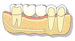
Option 1: Dental Bridges – The Traditional Fixed Solution

What is a dental bridge?

A dental bridge uses two or more adjacent teeth for support and is commonly used to replace one or more missing teeth. The bridge literally “bridges” the gap where teeth are missing.


Components of a Dental Bridge

Abutment Teeth (Anchor Teeth)

The supporting structure:

✓ Natural teeth on either side of the gap ✓ Serve as anchors for the bridge ✓ Must be structurally sound and healthy ✓ Provide stability and retention for the replacement


Pontic (Replacement Tooth/Teeth)

The artificial tooth:

✓ Replaces the missing tooth or teeth ✓ Attached to abutment crowns as integral part of bridge ✓ Designed to match your natural teeth in size, shape, and color ✓ Restores function and appearance


The Dental Bridge Procedure

What to expect:

Step 1: Preparation of Abutment Teeth

The supporting teeth must be filed down (reduced) to accommodate the bridge:

  • Remove outer layer of enamel
  • Reshape tooth to smaller dimensions
  • Create space for crown portion of bridge
  • Ensure proper fit and alignment

This preparation is irreversible—the teeth are permanently altered.


Step 2: Impressions and Temporary Bridge

✓ Precise impressions of prepared teeth ✓ Record of your bite relationship ✓ Temporary bridge placed while permanent is fabricated ✓ Laboratory creates custom bridge (typically 2-3 weeks)


Step 3: Final Bridge Placement

✓ Remove temporary bridge ✓ Try-in and verify fit of permanent bridge ✓ Check bite, appearance, and comfort ✓ Cement bridge permanently in place ✓ Final adjustments and polishing


Bridge Material Options

Choices for Glen Iris patients:

Tooth-Colored Materials:

✓ All-ceramic/porcelain: Natural appearance, ideal for front teeth ✓ Porcelain-fused-to-metal (PFM): Strong with natural look, suitable for back teeth ✓ Zirconia: Extremely strong ceramic, works for front and back teeth

Metal Materials:

✓ Gold alloys: Extremely durable, excellent for back teeth ✓ Base metal alloys: Strong and resistant to corrosion

Material selection considerations:

  • Location in mouth (visible vs. back teeth)
  • Chewing forces on replacement area
  • Aesthetic preferences
  • Budget considerations

Dr. Kaufman discusses which materials best suit your specific situation.


Advantages of Dental Bridges

Why bridges work well:

✓ Fixed in place (doesn’t come out like dentures) ✓ Relatively quick treatment (completed in 2-3 weeks typically) ✓ Restores function immediately once placed ✓ Natural appearance with modern materials ✓ No surgery required (unlike implants) ✓ Proven longevity (10-15+ years with proper care) ✓ Familiar technology (dentists have extensive experience)


Disadvantages of Dental Bridges

Important limitations:

⚠ Requires damaging healthy teeth (major drawback) ⚠ Abutment teeth weakened by reduction ⚠ Increased risk of decay and problems in supporting teeth ⚠ Doesn’t prevent bone loss under pontic ⚠ Cleaning challenges (special floss threaders needed) ⚠ If abutment tooth fails, entire bridge fails ⚠ Not ideal if adjacent teeth are perfectly healthy

The requirement to permanently alter healthy adjacent teeth is the primary concern Glen Iris patients have about bridges.


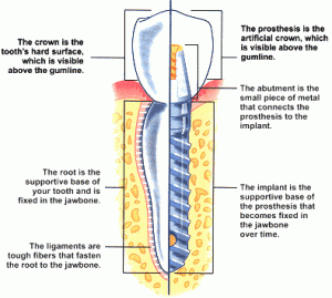
Option 2: Dental Implants – The Gold Standard Replacement

What are dental implants?

Dental implants are artificial tooth roots surgically placed into the jawbone. Once integrated with bone, they support replacement teeth such as crowns, bridges, or dentures.


Components of a Dental Implant

The Implant Fixture (Artificial Root)

Submerged in jawbone:

✓ Made of titanium or similar biocompatible materials ✓ Threaded post that integrates with bone (osseointegration) ✓ Replaces the missing tooth root ✓ Provides stable foundation for replacement tooth


The Abutment (Connector)

Middle component:

✓ Connects implant to crown ✓ Placed after healing period ✓ Custom-designed for each case


The Crown (Visible Tooth)

What you see:

✓ Custom-made to match natural teeth ✓ Attached to abutment ✓ Functions like natural tooth


The Dental Implant Process

Timeline and stages:

Phase 1: Implant Placement Surgery

✓ Surgical placement of titanium implant into jawbone ✓ Performed under local anesthesia (very comfortable) ✓ Implant is buried in bone beneath gum tissue ✓ Gum sutured closed over implant


Phase 2: Healing and Osseointegration

Critical integration period:

✓ Implant left undisturbed for 3-6 months ✓ Bone grows around implant (fuses with titanium) ✓ Establishes stable, permanent foundation ✓ Patient may wear temporary replacement during healing


Phase 3: Abutment Placement

✓ Minor procedure to expose healed implant ✓ Abutment attached to implant ✓ Gum tissue shaped around abutment ✓ Brief healing period (1-2 weeks)


Phase 4: Crown Fabrication and Placement

✓ Impressions taken for custom crown ✓ Crown designed to match natural teeth perfectly ✓ Crown attached to abutment ✓ Final adjustments for optimal function and appearance

Total timeline: Typically 4-8 months from start to finish.


Advantages of Dental Implants

Why implants are considered the best tooth replacement in Glen Iris:

✓ Most like natural tooth in function and feel ✓ Adjacent teeth not damaged or altered in any way ✓ Prevents bone loss (stimulates bone like natural root) ✓ Preserves facial structure and prevents aging appearance ✓ Independent (doesn’t rely on neighboring teeth) ✓ Excellent longevity (can last lifetime with proper care) ✓ Best chewing efficiency (closest to natural tooth) ✓ Doesn’t affect adjacent teeth (unlike bridges) ✓ Easy maintenance (brush and floss like natural teeth) ✓ No dietary restrictions once healed ✓ Highest patient satisfaction rates

The closest thing to a natural tooth:

For most Glen Iris patients, dental implants represent the gold standard tooth replacement option when feasible.


Disadvantages of Dental Implants

Considerations:

⚠ Requires adequate bone (bone grafting may be needed first) ⚠ Surgical procedure required (though routine and comfortable) ⚠ Longer treatment timeline (several months start to finish) ⚠ Higher initial investment (though excellent long-term value) ⚠ Medical conditions may contraindicate (uncontrolled diabetes, recent radiation, etc.) ⚠ Requires commitment to excellent oral hygiene

Despite these considerations, most Glen Iris patients find implants worth the investment.


Option 3: Dentures – The Removable Alternative

What are dentures?

Dentures are removable appliances that replace missing teeth and surrounding bone structure lost after extraction. Unlike bridges and implants, dentures can be removed and replaced by the wearer at will.


Types of Dentures

Full Dentures (Complete Dentures)

Replace all teeth in upper or lower arch (or both):

✓ Custom-made acrylic base matching gum color ✓ Artificial teeth set in base ✓ Relies on suction (upper) or gravity/muscles (lower) for retention ✓ Used when no natural teeth remain


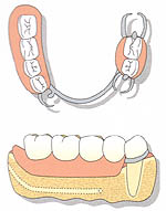
Partial Dentures

Replace some teeth while natural teeth remain:

✓ Replacement teeth attached to gum-colored base ✓ Metal or plastic framework ✓ Clasps hook onto remaining natural teeth for retention ✓ Removable for cleaning


How Dentures Function

Support and retention:

Dentures are supported by:

  • Jawbone ridge (where teeth used to be)
  • Remaining natural teeth (for partial dentures)
  • Gum tissues
  • Weak adhesion (denture adhesive may be needed)

The fundamental difference:

Unlike bridges and implants that are permanently fixed in your mouth, dentures are removable—which creates both advantages and significant disadvantages.


Advantages of Dentures

Limited benefits:

✓ Non-invasive (no surgery or tooth preparation) ✓ Can replace many teeth simultaneously ✓ Removable for cleaning (easier hygiene in some ways) ✓ Can be repaired or adjusted relatively easily ✓ Immediate solution (can be fabricated quickly)

Primary advantage: Dentures are typically less expensive initially than fixed bridges or implants.


Disadvantages of Dentures

Significant limitations Glen Iris patients experience:


1. Stability and Retention Problems

⚠ Can come out if you laugh, sneeze, or cough ⚠ Move while eating (uncomfortable, embarrassing) ⚠ Shift during speaking (affecting speech clarity) ⚠ Require adhesive for many wearers (messy, inconvenient) ⚠ Lower dentures particularly unstable (no suction, just gravity)


2. Functional Limitations

⚠ Significantly reduced chewing efficiency (approximately 25-30% of natural teeth) ⚠ Dietary restrictions necessary (avoid hard, sticky, or tough foods) ⚠ Cannot bite into foods with front teeth (apples, corn on cob) ⚠ Learning curve required for eating and speaking ⚠ Constant awareness of artificial appliance


3. Hygiene and Maintenance Challenges

⚠ Must remove and clean after every meal ⚠ Food traps underneath (uncomfortable, requires cleaning) ⚠ Sore spots develop from pressure points ⚠ Requires overnight removal (soak in cleaning solution) ⚠ Regular adjustments needed as fit changes


4. Progressive Bone Loss

The most serious long-term problem:

⚠ Dentures press on jawbone during function ⚠ This loading accelerates bone loss (opposite of natural tooth stimulation) ⚠ As bone shrinks, denture fit worsens progressively ⚠ Facial collapse occurs over years (sunken appearance, premature aging) ⚠ Remaining teeth become loose from denture pressure (partial dentures) ⚠ Eventually bone loss makes denture wearing extremely difficult

The vicious cycle:

More denture wear → More bone loss → Worse fit → More pressure → Accelerated bone loss

This is why Glen Iris patients wearing dentures for many years often have severely resorbed jawbones and aged facial appearance.


5. Impact on Quality of Life

⚠ Self-consciousness about dentures falling out ⚠ Social anxiety (worry about eating in public, speaking) ⚠ Reduced confidence ⚠ Constant inconvenience of removal and cleaning ⚠ Limitation of spontaneity (careful about what you eat, when, where)


Comparing Your Tooth Replacement Options in Glen Iris

Decision matrix:

Factor Dental Bridge Dental Implant Denture
Permanence Fixed Fixed Removable
Adjacent teeth Requires alteration Not affected May damage over time
Bone preservation No Yes No (accelerates loss)
Function Excellent Excellent Limited
Longevity 10-15+ years Lifetime potential 5-7 years average
Maintenance Moderate Easy High
Initial investment Moderate Higher Lower
Long-term value Good Excellent Poor
Treatment time 2-3 weeks 4-8 months 4-6 weeks
Surgery required No Yes No

Dr. Kaufman’s Recommendations for Glen Iris Patients

Personalized treatment planning:

The “best” tooth replacement option in Glen Iris depends on your individual situation:


When Dental Implants Are Ideal:

✓ Single tooth loss with adequate bone ✓ Adjacent teeth are healthy (shouldn’t be altered) ✓ Desire for most natural replacement ✓ Good overall health ✓ Commitment to excellent hygiene ✓ Willingness to invest in best long-term solution

Dr. Kaufman’s perspective: When feasible, implants offer the best outcomes for most Glen Iris patients.


When Dental Bridges Are Appropriate:

✓ Adjacent teeth already have large fillings or crowns (will need crowns anyway) ✓ Adequate bone isn’t present for implant (and grafting not desired) ✓ Medical conditions contraindicate surgery ✓ Shorter treatment timeline needed ✓ Budget doesn’t allow implant treatment


When Dentures May Be Necessary:

✓ Multiple teeth missing (full-arch replacement) ✓ Insufficient bone for implants (extensive grafting required) ✓ Medical conditions preventing surgery ✓ Immediate budget constraints ✓ Temporary solution while planning definitive treatment

Modern alternative: Implant-supported dentures combine denture convenience with implant stability—excellent middle-ground option.


Making Your Decision

Factors to consider:

✓ Long-term goals for your dental health ✓ Overall health status and medical conditions ✓ Adjacent teeth condition (healthy vs. already compromised) ✓ Bone quality and quantity available ✓ Timeline preferences (quick vs. optimal long-term) ✓ Budget and investment capacity ✓ Functional expectations (chewing, appearance, comfort) ✓ Willingness to undergo surgery (for implants)

Dr. Kaufman provides honest assessment of all options, helping you make the informed decision that’s right for your unique circumstances.


Comprehensive Tooth Replacement in Glen Iris

Dr. Kaufman and the team at Tooronga Family Dentistry provide all tooth replacement options with expertise and personalized care:

✓ Thorough evaluation of your situation ✓ Discussion of all appropriate options ✓ Honest pros and cons for each approach ✓ Digital imaging and treatment planning ✓ High-quality materials and craftsmanship ✓ Ongoing maintenance and support

Our tooth replacement services include:

✓ Single and multiple tooth dental implants ✓ Custom dental bridges (all materials) ✓ Partial and full dentures ✓ Implant-supported dentures ✓ Bone grafting when needed ✓ Comprehensive treatment planning

Schedule your tooth replacement consultation:

  • Phone: 9822 7006
  • Services: Dental implants, bridges, dentures, comprehensive tooth replacement planning, bone grafting
  • Location: Serving Glen Iris, Malvern, Ashburton, Camberwell, and surrounding Melbourne communities

Don’t let tooth loss compromise your oral health, appearance, and quality of life. Whether you’ve recently lost a tooth or have been living with gaps for years, tooth replacement options in Glen Iris can restore your smile, function, and confidence.

👉Call or book online Tooronga Family Dentistry on (03) 9822 7006  to learn more about tooth replacement options or schedule your comprehensive evaluation.

Every tooth matters. Replace what’s missing. Restore your complete smile.

Warning Signs of Gum Disease: Expert Treatment in Glen Iris

Posted on 09.7.15

Do your gums feel itchy or uncomfortable? Does eating cause gum pain? Do you notice bleeding when you brush your teeth or bite into an apple? These symptoms may indicate gum disease (also called periodontal disease)—a group of chronic conditions affecting the tissues supporting your teeth. At Tooronga Family Dentistry, Dr. Kaufman wants every Glen Iris patient to recognize the warning signs of gum disease and understand that early gum disease treatment in Glen Iris can prevent serious consequences including tooth loss and jawbone destruction.

The insidious nature of gum disease is that it’s extremely common yet usually goes unnoticed because it causes little or no pain in early stages. Understanding the symptoms and seeking prompt treatment protects your smile for life.


Why Gum Disease Goes Unnoticed

The silent progression:

Gum disease is one of the most prevalent health conditions worldwide, yet many Glen Iris patients remain unaware they have it because:

⚠ Early stages are painless (no alarm bells ring) ⚠ Symptoms seem minor (easily dismissed as “normal”) ⚠ Progression is gradual (changes happen slowly over months/years) ⚠ Many people normalize bleeding gums (assuming it’s typical) ⚠ Dental visits are infrequent (years pass without professional evaluation)

The dangerous assumption:

Many Glen Iris residents tend to disregard:

  • An odd painful feeling in the mouth
  • Occasional bleeding when brushing
  • Blood when biting into hard foods
  • Persistent bad breath
  • Slight gum swelling or redness

These are NOT normal—they’re warning signs requiring professional gum disease treatment.


The Two Stages of Gum Disease

Understanding the progression:

Gum disease exists on a continuum, with two distinct stages that Glen Iris patients should recognize:


Stage 1: Gingivitis (Reversible Gum Inflammation)

What is gingivitis?

Gingivitis represents mild to moderate inflammation of your gums caused by bacterial plaque accumulation around teeth.

Key characteristics:

✓ Affects gum tissue only (not yet involving bone) ✓ Reversible with proper treatment and home care ✓ Very common (affects majority of adults at some point) ✓ Often painless (making it easy to ignore) ✓ Early warning stage before serious damage occurs

The critical window:

Gingivitis is your opportunity to prevent progression. With proper gum disease treatment in Glen Iris, gingivitis can be completely reversed, restoring gums to healthy condition.


Stage 2: Periodontitis (Advanced Gum Disease)

What is periodontitis?

Without proper dental care, gingivitis progresses into periodontitis—a much more serious condition affecting not just gums but the supporting structures of teeth.

Destructive progression:

⚠ Inflammation spreads deeper into supporting tissues ⚠ Periodontal ligament breaks down (connects tooth to bone) ⚠ Jawbone tissue is destroyed (irreversible bone loss) ⚠ Gum pockets deepen (creating bacteria havens) ⚠ Teeth become loose from lost support ⚠ Tooth loss occurs in advanced stages

The irreversible reality:

Unlike gingivitis, periodontitis causes permanent damage. While gum disease treatment can halt progression and stabilize the condition, bone and attachment loss cannot be fully regenerated (though some improvement is possible with advanced therapies).

This is why early intervention at the gingivitis stage is so crucial for Glen Iris patients.


Comprehensive Warning Signs: Do You Have Gum Disease?

Self-assessment checklist:

Contact Tooronga Family Dentistry if you experience any of these symptoms:


Bleeding Gums (The Primary Warning Sign)

🚨 Gums bleed easily when brushing or flossing 🚨 Blood appears when chewing hard or crunchy foods (apples, carrots, nuts) 🚨 Spontaneous bleeding without any apparent reason 🚨 Pink toothbrush or blood in sink after brushing

Critical understanding: Healthy gums do NOT bleed. If your gums bleed, even occasionally, you have inflammation requiring professional evaluation and gum disease treatment in Glen Iris.

Many Glen Iris patients mistakenly believe bleeding gums are “normal” or caused by “brushing too hard.” This dangerous misconception delays treatment, allowing gingivitis to progress to periodontitis.


Gum Tenderness and Sensitivity

🚨 Gums are tender to touch (hurt when you apply pressure) 🚨 Discomfort when eating certain foods 🚨 Sensitivity along gum line (not tooth sensitivity) 🚨 Soreness that comes and goes

What it indicates: Active inflammation in gum tissues from bacterial infection.


Visible Gum Changes

🚨 Red or dark red gums (healthy gums are pale pink) 🚨 Swollen or puffy gums (healthy gums lie flat against teeth) 🚨 Gums separating from teeth (creating visible gaps or spaces) 🚨 Receding gums (teeth appear longer than before) 🚨 Shiny appearance (instead of stippled texture of healthy gums)

Visual comparison: Look at your gums in the mirror. Healthy gums are pale pink, firm, and fit snugly around each tooth. Diseased gums appear red, swollen, and pull away from teeth.


Persistent Bad Breath (Halitosis)

🚨 Chronic bad breath that doesn’t improve with brushing 🚨 Foul taste in mouth throughout the day 🚨 Metallic or unpleasant taste that persists 🚨 Bad breath returns quickly after brushing or mouthwash

The bacterial connection: Gum disease bacteria produce sulfur compounds causing characteristic bad breath. If persistent halitosis doesn’t resolve with improved oral hygiene, gum disease is a likely cause.


Pain and Discomfort

🚨 Hurts when you chew (especially on certain teeth) 🚨 Pain when food gets caught between teeth or in gum pockets 🚨 Aching or throbbing in gum areas 🚨 Discomfort that worsens over time

What it indicates: Advanced inflammation or infection in deeper periodontal structures.


Tooth Mobility and Position Changes

🚨 Teeth starting to feel loose (slight movement when touched) 🚨 Teeth shifting position (spaces developing, teeth tilting) 🚨 Changes in your bite (teeth fit together differently) 🚨 Teeth drifting forward (especially lower front teeth)

Critical warning: Tooth mobility indicates significant bone loss from periodontitis. This is an advanced stage requiring immediate professional gum disease treatment in Glen Iris.


Additional Warning Signs

🚨 Pus between teeth and gums (visible drainage) 🚨 Gum abscesses (painful, swollen bumps on gums) 🚨 Teeth appearing longer (from gum recession) 🚨 Changes in how partial dentures fit 🚨 New spaces developing between teeth


Dr. Kaufman’s Comprehensive Gum Disease Examination

Professional diagnosis:

As part of every comprehensive examination at Tooronga Family Dentistry, Dr. Kaufman dedicates significant time to thoroughly evaluating your gum health. Early detection allows for simpler, more effective gum disease treatment.


Periodontal Probing (Measuring Gum Pockets)

The essential diagnostic tool:

Dr. Kaufman measures the spaces around your teeth using a periodontal probe—a small, ruler-like instrument with millimeter markings.

How probing works:

  1. Probe is gently inserted between tooth and gum
  2. Depth of the pocket is measured in millimeters
  3. Measurements taken at six points around each tooth
  4. Bleeding on probing is noted (indicates active inflammation)
  5. Results recorded for monitoring over time

Understanding the measurements:

  • 1-3mm depth: Healthy (normal sulcus depth)
  • 4mm depth: Early gum disease (gingivitis or early periodontitis)
  • 5-6mm depth: Moderate periodontitis (bone loss present)
  • 7mm+ depth: Advanced periodontitis (significant bone destruction)

Larger-than-normal measurements or gaps between your teeth and gums indicate gingivitis or periodontitis requiring treatment.


Visual Examination

What Dr. Kaufman assesses:

✓ Gum color (healthy pink vs. inflamed red) ✓ Gum texture (firm and stippled vs. smooth and puffy) ✓ Gum position (attached vs. receding) ✓ Bleeding tendency (healthy vs. easy bleeding) ✓ Plaque and tartar accumulation ✓ Visible pus or infection signs


Dental X-rays (Radiographic Evaluation)

Revealing hidden bone loss:

Dr. Kaufman may take dental X-rays to evaluate bone tissue around teeth. X-rays reveal:

✓ Bone level surrounding each tooth root ✓ Bone loss patterns (horizontal vs. vertical) ✓ Severity of destruction (how much bone remains) ✓ Tooth root anatomy and condition ✓ Areas requiring immediate attention

Why X-rays matter:

You cannot see bone with visual examination alone. X-rays provide critical information about periodontitis severity and guide treatment planning for Glen Iris patients.


Comprehensive Assessment

Additional evaluation includes:

✓ Tooth mobility testing (checking looseness) ✓ Bite analysis (how teeth come together) ✓ Risk factor assessment (smoking, diabetes, genetics, medications) ✓ Oral hygiene evaluation (effectiveness of home care) ✓ Medical history review (conditions affecting gum health)


Creating Your Personalized Treatment Plan

From diagnosis to recovery:

With comprehensive examination results, Dr. Kaufman creates a customized plan for your recovery from gum disease.


For Gingivitis (Early Stage):

Treatment typically includes:

✓ Professional deep cleaning (scaling to remove plaque and tartar) ✓ Oral hygiene instruction (proper brushing and flossing techniques) ✓ Antimicrobial rinses (if appropriate) ✓ More frequent cleanings initially (every 3-4 months) ✓ Re-evaluation after 4-6 weeks to confirm reversal

Expected outcome: Complete reversal to healthy gums with proper treatment and home care.


For Periodontitis (Advanced Stage):

Treatment may include:

✓ Scaling and root planing (deep cleaning below gum line) ✓ Antimicrobial therapy (antibiotics or antimicrobial agents) ✓ Periodontal maintenance (cleanings every 3-4 months) ✓ Surgical intervention (for advanced cases—flap surgery, bone grafting) ✓ Adjunctive therapies (laser treatment, locally delivered antibiotics) ✓ Ongoing monitoring (regular re-evaluation)

Expected outcome: Halt disease progression, stabilize condition, prevent tooth loss, preserve remaining bone.


Risk Factors Increasing Gum Disease Susceptibility

Glen Iris patients at higher risk:

Understanding your risk factors helps Dr. Kaufman develop more aggressive prevention and gum disease treatment protocols:

⚠ Smoking or tobacco use (dramatically increases risk and severity) ⚠ Diabetes (particularly if poorly controlled) ⚠ Genetic predisposition (family history of gum disease) ⚠ Hormonal changes (pregnancy, menopause) ⚠ Medications causing dry mouth (reduces protective saliva) ⚠ Stress (weakens immune response) ⚠ Poor nutrition (vitamin deficiencies, especially Vitamin C) ⚠ Immune system disorders (HIV/AIDS, cancer treatment) ⚠ Teeth grinding or clenching (traumatic forces on periodontal tissues) ⚠ Misaligned teeth (difficult to clean properly)

High-risk Glen Iris patients require more frequent monitoring and proactive treatment approaches.


The Systemic Health Connection

Beyond your mouth:

Recent research reveals gum disease isn’t just a dental problem—it affects your entire body:

Gum disease links to:

  • Heart disease and stroke
  • Diabetes complications
  • Respiratory infections
  • Rheumatoid arthritis
  • Pregnancy complications (preterm birth, low birth weight)
  • Alzheimer’s disease
  • Certain cancers

The mechanism: Bacteria and inflammatory chemicals from infected gums enter the bloodstream, triggering systemic inflammation and disease processes throughout the body.

Treating gum disease improves not just oral health but overall health for Glen Iris patients.


Prevention: Your Best Defense

Protecting against gum disease:

✓ Brush twice daily (minimum two minutes each time) ✓ Floss daily (removes plaque between teeth where brushing can’t reach) ✓ Use interdental brushes if you have spaces between teeth ✓ Professional cleanings every six months (more frequently if recommended) ✓ Don’t smoke (single biggest preventable risk factor) ✓ Manage diabetes (if applicable—keep blood sugar controlled) ✓ Eat nutritious diet (adequate vitamins and minerals) ✓ Stay hydrated (maintains healthy saliva flow) ✓ Manage stress (supports immune function)


When to Seek Gum Disease Treatment

Don’t delay if you experience:

🚨 Any bleeding from gums (with or without cause) 🚨 Gum tenderness, redness, or swelling 🚨 Persistent bad breath despite good hygiene 🚨 Gum recession or teeth appearing longer 🚨 Loose teeth or position changes 🚨 Pain when chewing 🚨 Pus or discharge from gums

The earlier, the better: Gingivitis caught and treated early reverses completely. Periodontitis caught early prevents severe bone loss and tooth loss.


Expert Gum Disease Treatment in Glen Iris

Dr. Kaufman and the team at Tooronga Family Dentistry provide comprehensive gum disease diagnosis and treatment for Glen Iris patients at all stages:

Our periodontal services include:

✓ Thorough periodontal examinations with probing and X-rays ✓ Professional cleanings and periodontal maintenance ✓ Scaling and root planing (deep cleaning) ✓ Antimicrobial therapies ✓ Personalized oral hygiene instruction ✓ Risk factor modification counseling ✓ Ongoing monitoring and re-evaluation ✓ Referral to periodontists for complex cases

Schedule your gum disease evaluation:

  • Phone: 9822 7006
  • Services: Comprehensive periodontal examination, gum disease treatment, preventive care, periodontal maintenance
  • Location: Serving Glen Iris, Malvern, Ashburton, Camberwell, and surrounding Melbourne communities

If you’re suffering from bleeding gums or any other warning signs of gum disease, don’t wait for the condition to worsen. Early professional gum disease treatment in Glen Iris can reverse gingivitis completely and prevent the irreversible damage of periodontitis.

👉Call or book online Tooronga Family Dentistry on (03) 9822 7006 to schedule your comprehensive gum health evaluation.

Bleeding gums aren’t normal. They’re a warning. Get the treatment you need today.

  • A Calm and Relaxing Experience

    Start your journey
  • Happy Smiles, Our Passion

    Children's Dentistry
  • Dentistry for the Whole Family

    Explore Services
  • Straight Teeth for a Healthy Smile

    Learn More
  • When To Start

    Book now
  • Aesthetic Dentistry

    Book now

Orthodontic braces

Clear Aligners

Free Child Orthodontic assessment

Book an appointment

Our recent posts

  • Smile Makeovers in Glen Iris — Composite Bonding, Veneers and Cosmetic Dentistry
  • Best Toothpaste for Sensitive Teeth in Australia
  • Gum Disease and Bad Breath — What’s the Connection?
  • Enamel erosion is permanent. Understanding what causes it is the first step to stopping it.
  • With so many whitening options available, how do you know which one actually delivers results — safely?

RSS Know your teeth

  • The Fountain of Dental Youth
    The reason cosmetic dentistry is experiencing a boom is that baby boomers want to preserve their youthful appearance.
  • What Color Is Your Smile?
    Food and drink, illness, injury, heredity or environmental factors can discolor teeth.
  • The Fountain of Dental Youth
    The reason cosmetic dentistry is experiencing a boom is that baby boomers want to preserve their youthful appearance.

Australian Dental Association
Australian Society For treatment of Periodontal Disease and Implants
Australian Society For Dental Sedation
Society for Dental Care of Children.
Society for Esthetic and Cosmetic Dentistry
Study Club of Excellent Dentistry

Any use of this site constitutes your agreement to the Terms and Conditions and Privacy Policy. Please click on the links to read them.

Copy rights Dental Idea Pty. Ltd

  • Sitemap

Webdesign, creation and maintenance by Dental Idea Pty. Ltd.