Tooronga Family Dentistry in Glen Iris

Family dental care in Glen Iris

Book Now

98227006
0450067475

Suite 1.02, 1 Crescent Rd., Glen Iris 3146
  • Home
  • About Us
    • Dr Daniel Kaufman – Experienced Dental Care Professional in Glen Iris
    • Why Visit Tooronga Family Dentistry in Glen Iris?
    • Frequently Asked Questions – Dentist Glen Iris
    • Our Practice in Glen Iris
    • State of the art materials and equipment in Glen Iris
    • Strict Infection Control at Tooronga Family Dentistry in Glen Iris
  • Services
    • Orthodontic braces
    • Straight Teeth with Clear aligners in Glen Iris
      • Invisalign Clear Aligners: A Discreet Alternative to Braces in Glen Iris
    • Wisdom tooth extraction
    • Fixing broken teeth.
    • Kids Dentist in Glen Iris
    • Healing bleeding Gums and bad breath
    • Mending teeth with a large cavities.
    • Teeth Whitening
    • Dealing with dental fear
    • Eliminating bad breath – Halitosis
    • Replacing missing teeth
    • Reinforcing fragile teeth in Glen Iris
    • Treating Molar and Incisor Hypomineralization
  • Contact
    • Emergency Contact
    • Book an Appointment Online
    • Ask Dr Kaufman a question
  • Patient information
    • Our Practice in Glen Iris
    • Blog
    • What is a Root canal treatment
    • Why remove decay from teeth
    • Oral hygiene in Glen Iris
    • Gum Disease During Pregnancy in Glen Iris: Causes, Symptoms, and Risks
    • Crowns and Bridges in Glen Iris
    • What to do for traumatic tooth injuries in Glen Iris
    • Tooth removal – Extractions
    • Child Dental Benefits Schedule
    • Professionally made mouthguard
    • Practice policies in Glen Iris
    • Privacy Policy
    • Terms and Conditions
    • Sitemap

Could a Mother’s Immunity Shape Her Child’s Lifelong Gum Health? New Research Says Yes

Posted on 07.20.26

New research from the Hebrew University of Jerusalem has uncovered something remarkable: the immune protection a mother gives her baby doesn’t just guard against infections in infancy. It may actually shape that child’s resistance to gum disease for the rest of their life.

At Tooronga Family Dentistry in Glen Iris, we’re always interested in research that deepens our understanding of oral health across the lifespan. This study offers a fascinating new perspective on where the roots of gum disease resistance—and vulnerability—truly begin.

A Groundbreaking Study on Oral Immunity

The research identified that mothers equip their children with two distinct types of immune protection: antibodies transferred during pregnancy, and antibodies delivered through breast milk. Remarkably, each pathway plays a completely different long-term role in oral health.

How Antibodies Before Birth Program the Immune System

The first pathway involves antibodies transferred during pregnancy, known as in utero-derived IgG. These antibodies travel to the neonatal salivary glands and are secreted into saliva from birth.

Their job is more sophisticated than simple defence. These antibodies essentially teach the developing immune system how to behave—remaining calm around friendly, harmless bacteria while staying alert to genuine threats.

In laboratory models, offspring without these prenatal antibodies showed hyperactive immune cells and higher bacterial loads in their gums and salivary glands. Crucially, this early disruption led to lasting consequences: these offspring were significantly more susceptible to periodontitis in adulthood, the destructive gum condition that causes inflammation and bone loss around the teeth.

Breast Milk Antibodies Build the Mouth’s Physical Barrier

While prenatal antibodies set the immune “tone,” antibodies delivered through breast milk serve an entirely different purpose. This second pathway supports the physical maturation of the oral epithelium—the protective lining of the mouth.

These postnatal antibodies help regulate the timing of what researchers call “barrier sealing,” ensuring the mouth’s protective lining becomes sturdy at exactly the right developmental moment. This process is highly sensitive to environment. When breast milk antibodies were absent, or when their effects were disrupted by antibiotics, the physical integrity of this protective oral barrier was compromised.

Targeting the Bacteria Behind Aggressive Gum Disease

Perhaps most striking is the discovery that maternal antibodies specifically recognise and bind to certain oral pathobionts, including bacteria from the Pasteurellaceae family. These are bacteria known to drive particularly aggressive forms of gum disease.

This suggests that a mother’s immune system is doing far more than offering generic protection. It appears to be actively targeting the specific bacterial threats most likely to cause serious periodontal disease later in life.

What This Could Mean for the Future of Gum Disease Prevention

These findings open an exciting door: maternal immunisation. By vaccinating mothers during pregnancy, researchers suggest it may be possible to enhance the specific antibodies passed to a child, effectively pre-programming their immune system to resist chronic oral infections decades before symptoms would ever appear.

In other words, the foundations of a healthy adult smile may be laid before a single tooth has even erupted.

Why This Research Matters for Oral Health at Every Age

While this study focuses on the earliest stages of life, it reinforces something we see clinically every day at our Glen Iris practice: gum disease has deep biological roots, and individual susceptibility varies enormously from person to person.

This research helps explain why some patients are naturally more resistant to periodontal disease than others, even with similar oral hygiene habits. It’s a reminder that gum health is shaped by far more than brushing and flossing alone—though those daily habits remain essential tools we can all control.

Protecting Gum Health Throughout Life

Regardless of the immune foundation we’re born with, consistent oral care remains the most powerful tool for preventing periodontal disease:

  • Brushing twice daily with fluoride toothpaste
  • Daily flossing to remove bacteria between teeth
  • Regular professional cleanings every six months
  • Prompt treatment of early gum inflammation before it progresses
  • Ongoing monitoring for those with a family history of gum disease

Understanding that susceptibility to gum disease may begin before birth doesn’t change our recommendations—it reinforces why proactive, consistent dental care matters at every stage of life.

Comprehensive Periodontal Care at Tooronga Family Dentistry

At our Glen Iris practice, we take a thorough approach to assessing and protecting gum health for patients of all ages and risk profiles. Our team stays current with emerging research like this Hebrew University study, ensuring our patients benefit from the latest understanding of oral health science.

Whether you have a family history of gum disease, are noticing early warning signs like bleeding gums, or simply want a comprehensive periodontal assessment, we’re here to help.

Schedule Your Gum Health Assessment in Glen Iris

If it’s been a while since your last check-up, or you have concerns about your gum health, Call or book online Tooronga Family Dentistry on (03) 9822 7006 so we can help you.

We proudly care for the smiles of the Glen Iris, Tooronga, Malvern, Ashburton, Camberwell  and Hawthorn East communities

Our experienced team will assess your periodontal health, identify any risk factors, and create a personalised prevention plan tailored to your needs.

Keeping Your Natural Teeth Could Add Years to Your Independent Life, Singapore Study Reveals

Posted on 07.20.26

A groundbreaking study is changing how we think about ageing and dental care. Researchers have discovered that maintaining more of your natural teeth is directly linked to more years of independent living.

At Tooronga Family Dentistry in Glen Iris, we’ve always believed that protecting your natural teeth matters for life. Now, compelling new research confirms it: keeping your teeth may help you stay independent, mobile, and self-sufficient well into your later years.

The Singapore Study: Groundbreaking Research on Teeth and Ageing

The study was led by the National Dental Centre Singapore (NDCS) and published in the Journal of Epidemiology and Community Health.

A First-of-Its-Kind Study

This research holds particular significance because:

  • It’s a study to estimate how the number of natural teeth impacts years of life lived without limitations in daily activities
  • The study also measured physical function, such as walking and climbing stairs
  • Data came from a nationally representative longitudinal survey of more than 3,000 Singaporeans aged 60 and above
  • It provides some of the strongest evidence yet connecting oral health directly to healthy ageing outcomes

What the Researchers Measured

The study assessed how tooth retention influences two critical measures of independent living:

  • Activities of Daily Living (ADLs): Essential self-care tasks including bathing, dressing, and eating
  • Physical Function: Mobility measures such as walking and climbing stairs

The Remarkable Findings: More Teeth, More Independent Years

Among older adults who did not use removable dental prostheses, those who retained 20-32 natural teeth experienced substantially longer periods of independent living compared to those with no natural teeth:

At Age 60:

  • More than 5 additional years without ADL limitations
  • More than 3 additional years without physical function limitations

At Age 70:

  • More than 4 additional years without ADL limitations
  • 2.5 additional years without physical function limitations

At Age 80:

  • More than 2 additional years without ADL limitations
  • Over 1 additional year without physical function limitations

The Role of Dentures and Dental Prostheses

The research  revealed important findings about removable dental prostheses (dentures) for those who have already experienced tooth loss.

Dentures Can Help Bridge the Gap

Among prosthesis users with secondary or higher education:

  • Those with 20-32 natural teeth experienced more years without ADL limitations
  • Those with 10-19 natural teeth experienced more years without physical function limitations
  • Both groups showed improvements compared to those with no natural teeth

What This Means If You’ve Already Lost Teeth

This is encouraging news for anyone who has experienced tooth loss:

  • Removable dental prostheses may partially mitigate the effects of tooth loss
  • Proper denture care and fit remain important for maintaining function
  • It’s never too late to improve your oral health situation
  • Working with your Glen Iris dentist on prosthetic solutions can still provide meaningful benefits

The Science Behind Teeth and Independence

Understanding why natural teeth are so closely connected to independent living helps explain these remarkable findings.

Chewing Function and Nutrition

Natural teeth allow for:

  • Efficient chewing of a wide variety of nutritious foods
  • Better digestion when food is properly broken down
  • Adequate intake of protein, fibre, and essential nutrients
  • Maintenance of healthy body weight and muscle mass

Poor chewing function from tooth loss often leads to:

  • Avoidance of nutritious but harder-to-chew foods like meat, fruits, and vegetables
  • Shift toward softer, often less nutritious processed foods
  • Nutritional deficiencies that can accelerate physical decline
  • Reduced muscle strength and mobility over time

The Connection to Physical Function

Nutrition and physical capability are closely linked:

  • Poor nutrition contributes to muscle loss (sarcopenia)
  • Reduced muscle mass affects mobility, balance, and strength
  • This creates a cycle where poor oral health leads to poor nutrition, which leads to reduced physical function
  • Maintaining natural teeth helps break this cycle before it begins

Systemic Health Connections

Beyond nutrition, oral health connects to overall health in several ways:

  • Gum disease creates chronic inflammation linked to numerous health conditions
  • Oral infections can affect cardiovascular health
  • Poor oral health has been linked to cognitive decline
  • Chronic dental pain can reduce activity levels and social engagement

Protecting Your Natural Teeth: A Lifelong Investment

Essential Daily Oral Hygiene

Brushing Properly:

  • Brush twice daily with fluoride toothpaste
  • Spend at least two minutes each time
  • Use gentle, circular motions
  • Don’t forget your tongue and gum line
  • Replace your toothbrush every 3-4 months

Flossing Daily:

  • Remove plaque and food particles between teeth
  • Prevent gum disease that leads to tooth loss
  • Use proper technique, curving floss around each tooth
  • Consider water flossers if traditional floss is difficult

Professional Dental Care Throughout Life

Routine Check-ups:

  • Visit every six months for examination and cleaning
  • Early detection of problems prevents tooth loss
  • Professional cleaning removes plaque and tartar buildup
  • X-rays can identify hidden issues before they become serious

Gum Disease Prevention and Treatment:

  • Periodontal disease is a leading cause of tooth loss in adults
  • Early treatment can reverse gingivitis and prevent progression
  • Deep cleaning procedures address established gum disease
  • Ongoing maintenance prevents recurrence

Restorative Care When Needed:

  • Prompt filling of cavities prevents further decay
  • Root canal treatment can save teeth that might otherwise require extraction
  • Crowns protect weakened teeth from fracture
  • Addressing problems early preserves more of your natural tooth structure

Special Considerations for Ageing Adults

Managing Dry Mouth:

  • Many medications cause reduced saliva production
  • Dry mouth increases cavity and gum disease risk
  • Stay hydrated and consider saliva substitutes if needed

Addressing Gum Recession:

  • Gums naturally recede somewhat with age
  • Exposed roots are more vulnerable to decay
  • Special toothpastes can help with root sensitivity

Maintaining Mobility for Home Care:

  • Arthritis or reduced dexterity can affect brushing ability
  • Electric toothbrushes can help those with limited hand mobility

Preventing Tooth Loss: It’s Never Too Early or Too Late

If You Still Have All Your Natural Teeth

  • Maintain excellent daily oral hygiene
  • Don’t skip dental check-ups
  • Address any dental problems promptly
  • Consider this an investment in your future mobility and independence

If You’ve Lost Some Teeth

  • Protect the teeth you still have with excellent care
  • Discuss tooth replacement options for missing teeth
  • Consider dental implants, which function like natural teeth
  • Well-fitted dentures can help bridge nutritional and functional gaps

If You’ve Lost All Your Natural Teeth

  • Properly fitted dentures can partially mitigate functional limitations
  • Dental implants can support more stable, functional prostheses
  • Regular denture maintenance and adjustment remains important

Investing in Your Future Independence: Start Today

Take These Steps Today

  1. Schedule a comprehensive dental check-up if you haven’t had one recently
  2. Address any existing dental problems before they lead to tooth loss
  3. Commit to excellent daily oral hygiene to protect your remaining natural teeth
  4. Discuss your long-term oral health plan with your Glen Iris dentist
  5. If you’ve experienced tooth loss, explore your options for prosthetic solutions

Why Choose Tooronga Family Dentistry for Lifelong Oral Health

Comprehensive Preventive Care

  • Regular examinations to catch problems early
  • Professional cleanings to prevent gum disease
  • Personalised prevention plans based on your individual risk factors

Restorative Expertise

  • Prompt treatment of cavities to prevent tooth loss
  • Root canal therapy to save teeth that might otherwise be extracted
  • Crown and bridge work to protect and restore damaged teeth

Tooth Replacement Solutions

  • Dental implants for stable, natural-feeling replacement teeth
  • Well-fitted dentures for those who have experienced tooth loss
  • Bridges to replace missing teeth and maintain function

Protect Your Future Independence: Call or book online Tooronga Family Dentistry on (03) 9822 7006 to schedule your appointment and take a meaningful step toward protecting your future independence.

  • Location: Conveniently located in Glen Iris, Melbourne
  • Serving: Glen Iris, Toorak, Malvern, Camberwell, Ashburton, and surrounding suburbs

Our experienced team will:

  • Conduct a comprehensive evaluation of your current oral health
  • Identify any risk factors for future tooth loss
  • Create a personalised plan to protect your natural teeth
  • Discuss restoration or replacement options if needed

Is It Too Late to Straighten My Teeth?

Posted on 06.30.26

The short answer is no. The longer answer explains why adult orthodontics is one of the fastest-growing areas of dentistry — and why age is far less of a barrier than most people assume.

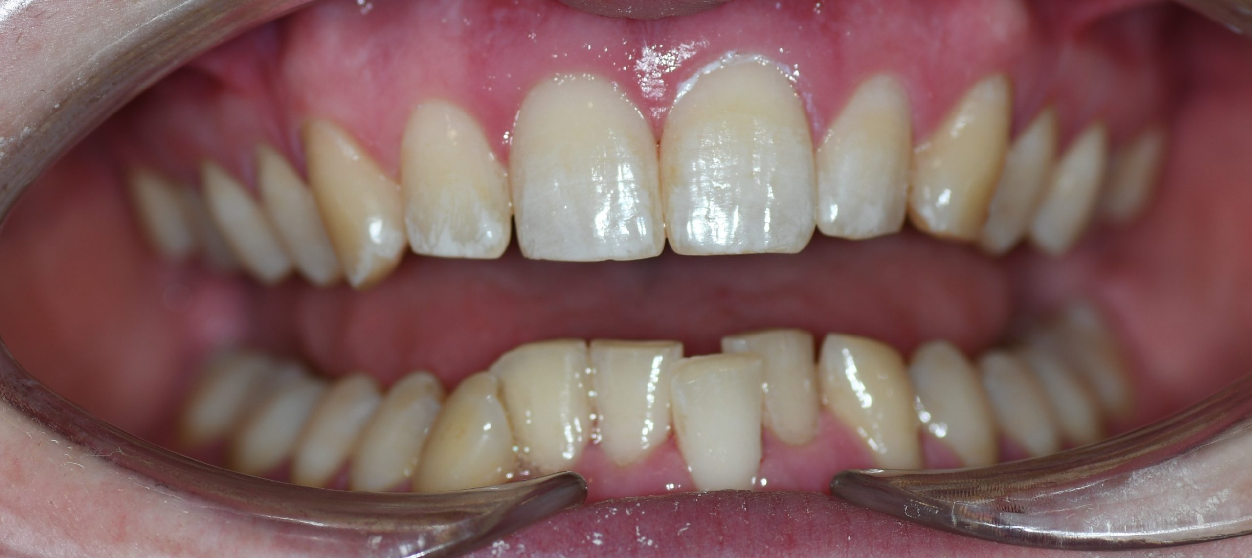
A significant proportion of adults in Glen Iris, Malvern, Hawthorn and Ashburton who attend Tooronga Family Dentistry have crowded, spaced, or misaligned teeth they have lived with for decades — often because they missed orthodontic treatment as children, had treatment that relapsed, or simply assumed that straightening teeth as an adult was not realistic. All three assumptions are worth examining.

Can teeth be moved at any age?

Yes — with one important qualification. Teeth move in response to sustained, gentle force applied to the periodontal ligament — the fibrous tissue suspending each tooth in its socket. This biological process does not have an age limit. Bone remodeling — the process by which bone dissolves on the pressure side of tooth movement and deposits on the tension side — occurs throughout life, albeit more slowly in older patients than in adolescents.

The prerequisite for orthodontic tooth movement is not age — it is adequate bone support and periodontal health. Teeth with healthy gums and sufficient bone can be moved successfully at 40, 50, 60 or beyond. Teeth compromised by significant bone loss from untreated periodontal disease cannot be moved safely until that disease is treated and stabilized.

Why adults seek orthodontic treatment

Adults pursue orthodontic treatment for a wider range of reasons than adolescents — and the motivations are often more considered and more specific.

  • Crowding that has worsened with age — lower front teeth in particular tend to crowd progressively throughout life
  • Relapsed orthodontic treatment from childhood — teeth that were straightened and then shifted after retainer use was discontinued
  • Spaces between teeth — diastema — that have become more noticeable or widened over time
  • Bite problems — deep bites, open bites, crossbite — that are causing wear, jaw discomfort or functional problems
  • Preparation for restorative work — straightening teeth before placing crowns, veneers or implants to achieve optimal positioning
  • Aesthetic motivation — a smile that has always been a source of self-consciousness and that the patient is finally ready to address

Orthodontic options for adults

The range of orthodontic options available to adults has expanded significantly over the past two decades. The choice between systems depends on the complexity of tooth movement required, aesthetic preferences, lifestyle considerations, and budget.

Clear aligner therapy — Invisalign and equivalents

The most popular orthodontic option for adults seeking treatment today. A series of custom-fabricated clear plastic aligners — each worn for one to two weeks — progressively moves teeth toward the planned final position. Aligners are removable for eating, brushing and flossing, virtually invisible when worn, and do not require the dietary restrictions associated with fixed braces.

Invisalign and its equivalents are highly effective for mild to moderate crowding, spacing, and many bite discrepancies. Complex tooth movements — significant rotations, large vertical movements, severe skeletal discrepancies — have historically been more challenging with aligners than with fixed appliances, though advances in aligner technology and the use of attachments have substantially closed this gap.

The critical variable with aligner therapy is compliance. Aligners must be worn for 20 to 22 hours per day to achieve planned tooth movement on schedule. Patients who remove aligners frequently or for extended periods will experience delayed or incomplete results. Adults who are motivated and disciplined about wear time achieve excellent outcomes; those who struggle with compliance may be better suited to fixed appliances.

Fixed ceramic braces

Tooth-colored ceramic brackets bonded to the teeth — functionally identical to conventional metal braces but significantly less visible. A good option for patients who require complex tooth movements that are better managed with fixed appliances, but who want a more aesthetic appearance than metal brackets provide. Less visible than metal braces, more visible than aligners.

Fixed metal braces

The most versatile and mechanically capable orthodontic option — capable of achieving complex tooth movements that push the limits of aligner therapy. Less aesthetically appealing than clear options but typically more cost-effective and unambiguously effective for complex cases. Some adults choose metal braces without hesitation — particularly where treatment complexity makes them the clinically superior option.

Retainers — fixed and removable

For very minor tooth movement — a single rotated tooth, a small space, minor post-treatment relapse — a simple removable retainer or a fixed bonded retainer placed behind the teeth may achieve the desired result without full orthodontic treatment. We assess whether this simpler approach is appropriate at consultation.

What adult orthodontics cannot do

Orthodontics moves teeth within the jaw. It does not change the underlying skeletal relationship between the upper and lower jaws. Significant skeletal discrepancies — a substantially receded lower jaw, a severe underbite, a pronounced open bite of skeletal origin — may require orthognathic surgery in combination with orthodontics to achieve a stable functional and aesthetic result. These cases require specialist orthodontic assessment and surgical referral.

Periodontal health is needed

This point warrants repetition. Active gum disease is an absolute contraindication to orthodontic tooth movement. Moving teeth through infected, inflamed bone accelerates bone loss and can cause irreversible damage. Before any orthodontic treatment is commenced we ensure gum health is assessed, any active disease is treated and stabilized, and a maintenance programm is in place to monitor periodontal health throughout treatment.

For adults with a history of gum disease who want orthodontic treatment, this is not a reason to decline treatment — it is a reason to treat the gum disease first and then proceed with appropriate monitoring.

What about existing dental work?

Crowns, bridges, implants and veneers all require consideration in orthodontic planning.

  • Crowns and veneers can be moved orthodontically — the underlying tooth root moves normally — though the restoration may require replacement after treatment if the shade or shape no longer matches adjacent teeth post-movement
  • Dental implants cannot be moved — they are fused directly to bone and have no periodontal ligament. Implant positions must be planned around the final orthodontic tooth positions, which is why orthodontics should ideally be completed before implant placement where both are planned
  • Bridges spanning a gap cannot be moved as a unit — the teeth supporting the bridge move independently, which can disrupt the bridge. Bridge-related planning requires careful assessment

How long does adult orthodontic treatment take?

Treatment duration varies with the complexity of movement required and the system used. Minor tooth movement — small spaces, mild crowding — may be completed in four to six months with aligners. Moderate cases typically require twelve to eighteen months. Complex cases may extend to twenty-four months or beyond.

Adult treatment generally takes slightly longer than equivalent treatment in adolescents — bone remodeling is slower with age. This is a modest difference in most cases and is rarely a clinically significant factor in treatment planning.

Retention — the phase that determines whether the result lasts

This is the most important thing most adults do not know about orthodontics: the result of orthodontic treatment is not permanent without retention. Teeth have an inherent tendency to return toward their pre-treatment positions — particularly lower front teeth — driven by the elastic recoil of the periodontal ligament and ongoing jaw development. Without sustained retention, relapse is the default outcome.

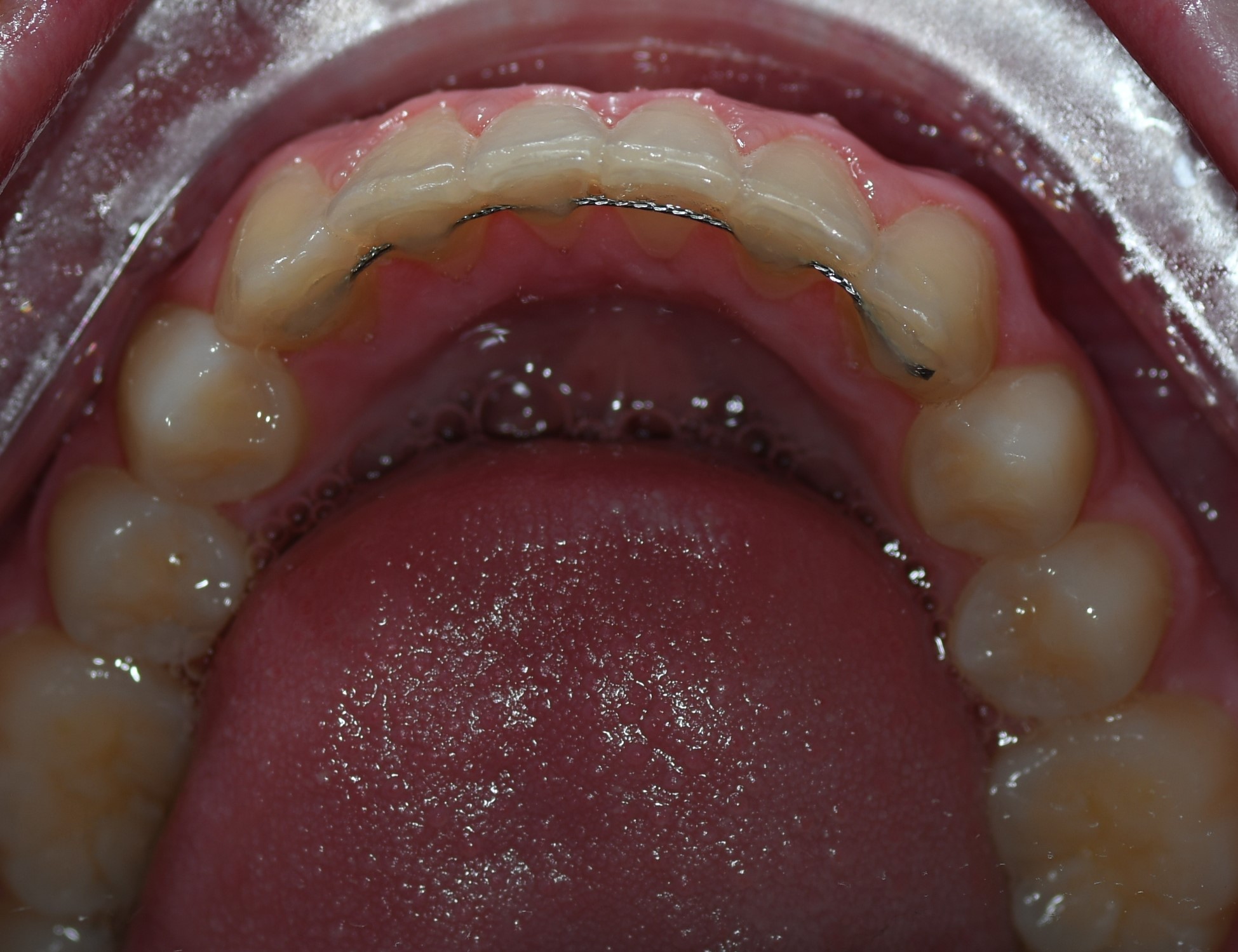
Modern retention protocols use a combination of fixed bonded retainers — thin wires bonded behind the upper and lower front teeth — and removable retainers worn nightly. Fixed retainers hold teeth in position continuously; removable retainers are used as a backup and for the back teeth not covered by the fixed wire.

Retention is a lifelong commitment for adults who want to maintain their result permanently. Removing retainers entirely after a period of years — as was common practice in older protocols — reliably produces relapse over time. We discuss this openly at the outset of treatment at Tooronga Family Dentistry so that expectations and commitment are realistic.

Starting the conversation at Tooronga Family Dentistry

Dr. Kaufman offers orthodontic consultations for adults across Glen Iris, Malvern, Hawthorn, Hawthorn East and Ashburton who are considering straightening their teeth. Our assessment covers the complexity of tooth movement required, the most appropriate treatment system, periodontal health assessment, any restorative considerations, realistic treatment duration and cost, and retention planning.

Once an assessment is done, Dr. Kaufman will present the condition and give honest advice about what is achievable, what the trade-offs are between different systems, and whether treatment is in your best clinical and aesthetic interest.

Thinking about straightening your teeth but assumed it was too late? It almost certainly isn’t. Call or book online Tooronga Family Dentistry on (03) 9822 7006  a consultation at Tooronga Family Dentistry — we see patients from Glen Iris, Malvern, Hawthorn, Hawthorn East and Ashburton and provide clear, honest advice about adult orthodontic options.

Gum Recession — Causes, Consequences and How It Is Fixed

Posted on 06.29.26

Gum recession is one of the most under-treated conditions in dentistry. Many people notice it, fewer understand its implications, and fewer still know that it can be corrected.

Receding gums — gums that have pulled away from the tooth surface, exposing the root beneath — affect a significant proportion of adults. The exposed root is not simply an aesthetic concern. It is a clinical problem with real consequences for tooth sensitivity, structural integrity, and long-term tooth survival. Understanding what causes recession, what happens if it is left untreated, and what can be done about it is the starting point for protecting teeth that are already showing signs of exposure.

What is gum recession?

The gum tissue — normally forms a tight seal around the neck of each tooth, protecting the root surface and underlying bone from bacteria, temperature, and mechanical trauma. When this tissue recedes — moves apically, away from the crown and toward the root — it exposes the cementum-covered root surface beneath.

Unlike enamel, which covers the crown, root cementum is thin, porous, and highly sensitive. Exposed root surfaces are immediately vulnerable to sensitivity, bacterial invasion, and physical abrasion. Over time, recession that is not treated progresses — slowly in most cases, rapidly in others — with each millimeter of further tissue loss compounding the clinical and aesthetic consequences.

What causes gum recession?

Recession rarely has a single cause. In most patients it is due to several contributing factors acting simultaneously or sequentially.

Toothbrush abrasion The most common cause of localized recession in otherwise periodontally healthy patients. Aggressive horizontal brushing with a medium or hard-bristled toothbrush physically damages the gum tissue over years of cumulative trauma. The pattern is characteristic — recession typically affects the outer surfaces of teeth most prominent in the arch, with a notch or groove at the gum-line of the affected teeth. The damage is slow and painless until the root is sufficiently exposed to cause sensitivity, by which point significant tissue has already been lost.

Periodontal disease Chronic periodontitis destroys the bone and connective tissue attachment supporting the teeth from below. As attachment is lost, the gum tissue follows — receding to the level of the remaining bone support. Recession from periodontal disease tends to be generalized rather than localized to one or two teeth, and is accompanied by the other features of active gum disease — bleeding, deep pockets, bone loss on X-ray.

Thin gum biotype The gum biotype — the inherent thickness and quality of gum tissue — varies significantly between individuals and is largely genetically determined. Patients with a thin, delicate gum biotype are significantly more susceptible to recession from any mechanical or inflammatory insult than those with thick, fibrous gum tissue. A thin biotype is not a pathological finding — it is an anatomical characteristic — but it means the threshold for recession is lower and the consequences of trauma or inflammation are more pronounced.

Orthodontic treatment Tooth movement outside the limits of the supporting alveolar bone — particularly moving teeth labially, toward the outer surface — can stretch thin gum tissue to the point of recession. This is a recognized risk of orthodontic treatment, particularly with clear aligner therapy where teeth may be moved beyond their natural bone envelope. Careful treatment planning and awareness of gingival biotype reduces but does not eliminate this risk.

High frenum attachments A frenum — the small band of tissue connecting the lip or cheek to the gum — that attaches high on the gum margin creates continuous tension that can pull gum tissue away from the tooth over time. A prominent mid-line frenum between the upper central incisors is a common contributor to recession at that specific site.

Bruxism and clenching The excessive forces generated by grinding and clenching create lateral stress at the cervical region of the tooth — the neck where crown meets root — that can contribute to both tooth flexure abrasion and gum recession. Bruxism-related recession tends to be accompanied by other signs of grinding — worn biting surfaces, tooth fractures, jaw muscle tenderness.

Lip and tongue piercings Chronic contact of jewellery against gum tissue creates persistent mechanical trauma that produces localized recession at the contact site. Lower central incisors are the most commonly affected teeth. The recession can be rapid and significant.

Tooth malposition A tooth that sits outside the natural arch — rotated, tipped, or positioned too far forward or backward — may have inadequate bone and gum coverage on one or more surfaces, predisposing it to recession regardless of oral hygiene.

What happens if gum recession is left untreated?

Recession is progressive. It does not stabilize spontaneously once it has begun — unless the cause is removed. Without addressing both the cause and the existing tissue loss, the following consequences develop over time:

Root sensitivity Exposed root cementum is acutely sensitive to temperature, touch, sweet and acidic stimuli. As recession progresses, sensitivity worsens. In advanced cases the sensitivity can be debilitating and affect dietary choices, oral hygiene compliance, and quality of life.

Root decay Root surfaces have no enamel protection. Exposed root cementum is significantly more vulnerable to decay than the enamel-covered crown. Root caries — cavities on the root surface — develop in the presence of plaque accumulation on exposed roots and can progress rapidly. Root decay in the context of recession requires filling and management of the underlying recession to prevent recurrence.

Notching and abrasion Continued brushing trauma on exposed root surfaces creates progressive abrasion — physical wearing away of the cementum and dentine — producing a visible V-shaped notch at the gumline. Once this notch has formed, the structural integrity of the tooth at that level is compromised.

Aesthetic concern Significant recession produces visible lengthening of the teeth — hence the expression “long in the tooth” — and an uneven, asymmetrical gumline. For anterior teeth particularly, this has a pronounced aesthetic impact that affects smile confidence.

Tooth loss In advanced cases where recession is associated with significant periodontal attachment loss, the teeth may become mobile and eventually lost. Recession alone — without bone loss — rarely leads directly to tooth loss, but it increases vulnerability to the conditions that do.

How gum recession is treated

Treatment has two components: addressing the cause to prevent further progression, and restoring the lost tissue where clinically indicated.

Addressing the cause

This is the essential first step. Restoring gum tissue without removing the cause of recession produces a result that will not last.

  • Brushing technique modification: Switching to a soft-bristled brush and retraining technique — gentle circular or modified Bass technique rather than horizontal scrubbing — is the single most important intervention for abrasion-related recession. We demonstrate correct technique at Tooronga Family Dentistry and recommend electric toothbrushes with pressure sensors for patients who consistently over-brush.
  • Periodontal treatment: Active gum disease must be treated before any surgical recession management. Scale and clean, root planing, and a structured maintenance program to arrest the inflammatory process driving bone and tissue loss.
  • Occlusal splint for bruxism: A custom nightguard reduces the excessive cervical forces associated with grinding and clenching.
  • Frenectomy: Surgical release of a high frenum attachment eliminates the tension contributing to localized recession at the affected site. Often performed in conjunction with gum grafting.
  • Orthodontic reassessment: Where tooth movement has contributed to recession, treatment planning modification may be required.

Gum grafting — restoring lost tissue

Where recession has produced clinically significant root exposure — causing sensitivity, aesthetic concern, or vulnerability to root decay — surgical tissue grafting is the definitive treatment for restoring gum coverage.

Connective tissue graft (subepithelial connective tissue graft) The gold standard procedure for recession coverage. Connective tissue is harvested from beneath the surface of the palate — leaving the overlying epithelium intact for faster palatal healing — and sutured into position over the exposed root at the recipient site. The graft becomes incorporated into the existing tissue, re-establishing a band of thick, keratinised gum tissue over the root.

This procedure produces highly predictable root coverage — complete coverage is achievable in carefully selected cases — and a natural-looking result that blends seamlessly with the surrounding tissue. It is performed under local anesthesia.

Free gingival graft A strip of tissue including the epithelial surface is harvested from the palate and placed at the recipient site. Produces a thicker band of keratinised tissue but with a less natural appearance than a connective tissue graft — the color match between graft and surrounding tissue is typically less precise. Used predominantly where the primary goal is increasing the width of attached gingiva rather than aesthetic root coverage.

Allografts and xenografts Tissue substitutes — processed human donor tissue or animal-derived collagen matrices — that eliminate the need for a palatal harvest site. Reduce post-operative discomfort and allow treatment of multiple sites simultaneously without the limitation of available donor tissue. Results are comparable to autogenous grafts in most studies, though some clinicians prefer autogenous tissue for optimal long-term outcomes.

Tunnel technique and modified approaches Minimally invasive alternatives to traditional grafting that use small incisions and tunnelling beneath the existing tissue to position the graft with minimal disruption to the blood supply. Associated with faster healing and excellent aesthetic outcomes in appropriate cases.

The pinhole surgical technique A minimally invasive approach using a small entry point through which existing gum tissue is repositioned coronally — toward the crown — over the exposed root, with a collagen matrix used to stabilize the new position. Less invasive than traditional grafting with faster healing, though long-term evidence is still accumulating compared to the extensive track record of connective tissue grafting.

Composite bonding for root coverage Where surgical grafting is not appropriate — due to patient preference, medical contraindications, or specific anatomical factors — tooth-coloured composite resin can be bonded to the exposed root surface to cover sensitivity, restore contour, and reduce vulnerability to root decay. This is not a tissue replacement — it does not restore biological tissue — but it is a clinically useful and aesthetically acceptable alternative in selected cases.

What to expect from gum grafting

Gum grafting is a well-established surgical procedure with a long clinical track record. Recovery typically involves mild to moderate discomfort for five to seven days, managed with ibuprofen and paracetamol. A soft diet is required for two to three weeks while the graft heals. Sutures are removed at one to two weeks. The full aesthetic result — complete colour and contour maturation — develops over three to six months as the graft matures and integrates.

The palatal harvest site, in autogenous grafting, is typically the primary source of post-operative discomfort — though patients are consistently surprised that it is less uncomfortable than anticipated. Modern surgical techniques and careful palatal management minimize this significantly.

Who performs gum grafting at Tooronga Family Dentistry?

We assess all patients with recession at Tooronga Family Dentistry and determine whether grafting is clinically indicated, what type of procedure is most appropriate, and whether treatment is best provided within our practice or referred to a periodontist — a specialist in gum tissue surgery — for complex multi-site grafting or cases with significant anatomical challenges.

We see patients from Glen Iris, Malvern, Hawthorn, Hawthorn East and Ashburton for gum recession assessment and management, and we provide a clear, honest recommendation based on clinical findings rather than a default toward surgery.

Is gum grafting worth it?

For patients with symptomatic recession — significant sensitivity, active root decay, progressive tissue loss — the answer is almost always yes. Restoring tissue coverage eliminates sensitivity, protects the root from further decay and abrasion, and halts the progression of tissue loss. The investment in time and cost is substantially less than the long-term cost of managing root decay, replacing worn or decayed root surfaces, or ultimately losing the tooth.

For patients with mild, stable recession and no symptoms, careful monitoring and cause elimination may be all that is required. We make this distinction clearly.

Noticing receding gums, root sensitivity, or longer-looking teeth? Book an assessment at Tooronga Family Dentistry — we see patients from Glen Iris, Malvern, Hawthorn, Hawthorn East and Ashburton and provide a thorough assessment of recession cause, severity, and the most appropriate treatment pathway.

How to Fix Yellow Teeth Fast — What Actually Works

Posted on 06.29.26

Yellow teeth are one of the most common cosmetic dental concerns — and one of the most fixable. The key is understanding what is causing the discoloration, because the fastest and most effective solution depends entirely on the answer.

Not all yellow teeth are the same. Surface staining from coffee responds differently to whitening than the deep yellowing caused by ageing dentine. Treating them identically produces inconsistent results — and in some cases, disappointment. A brief but accurate diagnosis changes everything.

Why teeth turn yellow

Tooth discoloration falls into two broad categories — extrinsic and intrinsic — and most people have a combination of both.

Extrinsic discoloration is staining on or near the outer surface of the tooth. The primary culprits are coffee, tea, red wine, tobacco, and certain foods with intense pigmentation. Tannins and chromogens in these substances bind to the protein pellicle coating the tooth surface and embed progressively into the outer enamel layer. This type of staining responds most rapidly and dramatically to professional cleaning and whitening.

Intrinsic discoloration originates within the tooth structure itself — in the dentine beneath the enamel. The most common cause is ageing: as enamel thins and becomes more translucent over time, the naturally yellow dentine beneath shows through more prominently. Trauma, certain medications taken during tooth development — tetracyclines in particular — fluorosis, and pulp changes following root canal treatment all cause intrinsic discoloration of varying degrees. Intrinsic discoloration requires whitening to address — cleaning alone will not change it.

The fastest options — ranked by speed of result

  1. In-chair professional whitening — same appointment, dramatic result For patients who want the fastest visible change, in-chair whitening is unmatched. A high-concentration hydrogen peroxide gel is applied to the teeth, often light-activated, over a single appointment of approximately 60 to 90 minutes. Most patients achieve a lift of four to eight shades in one session. You leave the appointment with visibly, dramatically whiter teeth.

At Tooronga Family Dentistry we offer in-chair whitening for patients across Glen Iris, Malvern, Hawthorn and Ashburton who have a specific event, occasion, or simply want the fastest possible result. A pre-whitening clean in the same visit — removing surface staining and calculus before the whitening agent is applied — maximises the outcome.

  1. Professional scale and polish — immediate surface stain removal For patients whose yellowing is primarily extrinsic — driven by coffee, tea, wine and tobacco — a professional scale and clean with air polishing removes surface staining instantly and reveals the natural tooth shade beneath. Many patients are genuinely surprised by how white their teeth actually are once the accumulated surface staining is professionally removed.

This is the fastest option for extrinsic staining and should precede any whitening treatment — whitening works more effectively on a clean tooth surface.

  1. Dentist-prescribed take-home whitening Custom trays made from impressions of your teeth, combined with professional-strength whitening gel. Results develop over ten to fourteen days of daily use and are comparable to in-chair whitening in final outcome. Not as fast as in-chair but significantly more effective than anything available over the counter, and the trays are reusable for maintenance top-ups indefinitely.
  2. Combined in-chair and take-home The most comprehensive approach. In-chair whitening delivers immediate impact; take-home consolidates and extends the result. The combination produces the most dramatic and the most durable outcome of any whitening approach.
  3. Over-the-counter whitening strips and kits Limited by Australian regulations to 6% hydrogen peroxide — significantly lower than professional concentrations. Can produce noticeable improvement in extrinsic staining over three to four weeks of consistent use. Slower and less dramatic than professional options but accessible without a dental appointment. A reasonable starting point for mild yellowing or maintenance between professional treatments.

What about whitening toothpastes?

Whitening toothpastes work through mild abrasion and low-concentration peroxide. They are effective at preventing new surface staining from accumulating and maintaining the result after professional whitening — but they do not lift established intrinsic yellowing. They are a maintenance tool, not a primary treatment.

What about charcoal products?

Heavily marketed, poorly evidenced, and potentially harmful. Charcoal toothpastes have no peer-reviewed clinical evidence demonstrating effective whitening beyond surface abrasion. Their abrasiveness can progressively thin enamel, ultimately making teeth appear more yellow — not less — as the underlying dentine shows through more prominently. Avoid them.

What about home remedies — lemon juice, baking soda, oil pulling?

Lemon juice and apple cider vinegar are erosive acids that dissolve enamel — using them on teeth is counterproductive and potentially damaging. Baking soda has mild abrasive action but no evidence for meaningful whitening beyond surface polishing. Oil pulling has no credible evidence for whitening. None of these approaches produces meaningful results, and some cause genuine harm.

What determines how white your teeth can get?

Whitening has a ceiling — it cannot make teeth whiter than their natural maximum. Several factors determine both the achievable result and how quickly it is reached:

  • Baseline shade — yellowish teeth typically respond better and faster than greyish teeth
  • Staining type — extrinsic staining responds fastest; intrinsic discolouration takes longer and varies by cause
  • Enamel thickness — thinner enamel from erosion or ageing affects both sensitivity and outcome
  • Existing restorations — fillings, crowns and veneers do not whiten; shade mismatches may require restoration replacement after whitening
  • Tetracycline staining — the most resistant to whitening; may require prolonged treatment or alternative cosmetic solutions such as veneers

When whitening is not the answer

For some patients, discolouration cannot be adequately addressed with whitening alone. Severe intrinsic staining, significant structural damage, or prominent restorations that cannot be whitened may be better addressed with composite bonding or porcelain veneers — covering the tooth surface with a matched, aesthetic material rather than attempting to lighten the underlying tooth.

We assess each patient’s situation individually at Tooronga Family Dentistry and recommend the approach most likely to achieve the result they are looking for — not the most expensive option or the most time-consuming one.

How to maintain your result and keep teeth white longer

The fastest way to lose a whitening result is to resume the habits that caused yellowing in the first place without any protective measures:

  • Rinse with water immediately after coffee, tea or red wine — dramatically reduces staining
  • Use a straw for cold coffee and iced tea to minimise contact with teeth
  • Use a low-abrasivity whitening maintenance toothpaste after treatment
  • Schedule a professional clean every six months — removing new surface staining before it embeds
  • Keep your take-home trays and use them for a top-up night every one to three months — this alone maintains results indefinitely for most patients
  • Avoid smoking — tobacco staining is among the most rapid and resistant to removal

Book a whitening consultation at Tooronga Family Dentistry

The fastest path to whiter teeth starts with an accurate assessment of what is causing the yellowing and a treatment plan matched to the result you want. We see patients from Glen Iris, Malvern, Hawthorn, Hawthorn East and Ashburton for whitening consultations, professional cleans and in-chair whitening — and we give honest, realistic advice about what is achievable and how quickly.

Ready to fix yellow teeth fast? Book a consultation at Tooronga Family Dentistry — we will identify the cause, recommend the right treatment, and deliver results you can se

Is It Safe to Whiten Your Teeth and How Long Does It Last?

Posted on 06.29.26

Teeth whitening is one of the most requested cosmetic treatments in Australia — and one of the most misunderstood. Here is what the evidence actually says about safety and longevity.

Two questions dominate every whitening consultation at Tooronga Family Dentistry: is it safe, and how long will it last? Both are entirely reasonable. Whitening is an elective cosmetic procedure — nobody should undergo it without understanding what it does to their teeth and what to realistically expect from the result.

Is teeth whitening safe?

The short answer is yes — professional teeth whitening performed or prescribed by a dentist is safe for enamel and the surrounding oral tissues when used at appropriate concentrations and for recommended durations. This conclusion is supported by decades of clinical research and an extensive track record in dental practice worldwide.

The longer answer requires understanding what whitening actually does — and where the genuine risks lie.

What whitening does to your teeth

Professional whitening uses hydrogen peroxide or carbamide peroxide as the active agent. These molecules are small enough to penetrate enamel and reach the dentine beneath, where they break apart the carbon double bonds in chromogen molecules — the organic compounds responsible for tooth discolouration. The result is lighter, less saturated colour without any removal or abrasion of tooth structure.

This is a fundamentally different mechanism from abrasive whitening — charcoal toothpastes, harsh whitening toothpastes, baking soda — which physically wear the enamel surface. Peroxide-based professional whitening does not remove enamel. Studies examining enamel hardness, surface morphology and mineral content before and after whitening at professional concentrations consistently find no clinically significant structural changes.

The genuine risks of whitening — and how they are managed

Tooth sensitivity The most common side effect of whitening, affecting a significant proportion of patients to some degree. Peroxide temporarily increases enamel permeability, allowing temperature and osmotic stimuli to reach dentinal tubules more readily. This produces a sharp, brief sensitivity — sometimes described as zingers — that is typically worst during treatment and in the 24 to 48 hours following. It resolves completely in the vast majority of patients.

At Tooronga Family Dentistry we manage whitening sensitivity proactively — assessing sensitivity risk before treatment, recommending desensitising toothpaste in the weeks prior, applying in-chair desensitising agents, and adjusting treatment duration or concentration where necessary. For patients with pre-existing sensitivity, a modified protocol minimises discomfort without compromising results.

Gum irritation Peroxide contact with gum tissue causes a reversible chemical irritation — whitening of the gum tissue, tenderness and mild inflammation. With professional in-chair whitening, a protective barrier is applied to the gums before treatment. With take-home whitening, custom-fitted trays are precision-made to cover teeth only, preventing gel pooling on gum tissue. Ill-fitting over-the-counter trays are the primary cause of significant gum irritation — a compelling reason to use professionally made trays rather than generic alternatives.

Overuse and unregulated products The risks of whitening are not inherent to the process — they are largely a product of misuse. High-concentration products used without professional supervision, over-the-counter products used excessively, and unregulated overseas products purchased online can cause enamel dehydration, increased porosity, and soft tissue damage. The Australian regulatory framework limiting over-the-counter products to 6% hydrogen peroxide exists for precisely this reason.

Pre-existing conditions requiring assessment before whitening

Whitening is not universally appropriate. Before recommending whitening we assess for:

  • Active decay — whitening an untreated cavity concentrates peroxide in a compromised area and must be avoided
  • Gum disease — inflamed, receding gums increase sensitivity risk and peroxide contact with exposed roots
  • Existing restorations — composite fillings, veneers and crowns do not respond to peroxide whitening; their shade remains fixed while surrounding teeth lighten, potentially creating a mismatch that requires restoration replacement
  • Enamel erosion — thinned enamel increases sensitivity risk and may not be suitable for whitening without prior remineralisation treatment
  • Pregnancy and breastfeeding — whitening is deferred during pregnancy as a precautionary measure; it is considered acceptable during breastfeeding but we discuss this individually

A thorough pre-whitening assessment is not a formality — it directly determines whether whitening is appropriate, which protocol is safest, and what results are realistically achievable.

What affects whitening results

Not all teeth whiten equally. Understanding what influences the outcome sets realistic expectations:

Type of staining Extrinsic staining — surface discolouration from coffee, tea, wine and tobacco — responds most readily and dramatically to whitening. Intrinsic staining — discolouration within the tooth structure itself — varies in its response depending on the cause. Age-related yellowing responds very well. Tetracycline staining — grey-brown banding from antibiotic exposure during tooth development — is among the most resistant to whitening and may require prolonged treatment or alternative cosmetic approaches.

Natural tooth shade Yellowish teeth typically whiten more effectively than greyish teeth. The baseline shade and underlying dentine colour influence both the achievable result and the time required.

Existing restorations As noted, composite and porcelain restorations do not whiten. Patients with prominent front fillings or crowns need to factor in the potential need for restoration replacement to match their new tooth shade.

Age Enamel becomes more permeable with age, which can actually make whitening more effective in older patients — though increased sensitivity risk must be considered.

How long does teeth whitening last?

This is the question with the most variable answer — because longevity depends almost entirely on lifestyle factors that differ significantly between individuals.

In-chair whitening results are immediately visible and typically dramatic — most patients achieve a lift of several shades in a single two-hour appointment. The initial result includes a degree of dehydration-related whitening that partially fades within the first one to two weeks as the teeth rehydrate. The stabilised result after rehydration is the true baseline from which longevity should be measured.

Take-home whitening produces results comparable to in-chair whitening over two to three weeks of daily use. The result develops more gradually but is no less effective in the final outcome.

How long results last:

For patients who avoid or minimise staining foods and drinks and maintain excellent oral hygiene, whitening results typically last one to two years before a top-up is needed. For patients who drink coffee, tea or red wine and do not use a whitening maintenance toothpaste, staining can return within six .

The most important factors determining longevity are:

  • Coffee, tea, red wine and tobacco consumption — the primary causes of staining recurrence; reducing frequency and rinsing with water after consumption significantly extends results
  • Oral hygiene — regular professional cleans remove surface staining before it embeds
  • Whitening toothpaste maintenance — low-abrasivity whitening toothpastes used after treatment help maintain results between professional top-ups
  • Top-up treatment — patients who use their take-home trays for one to two nights every three to six months maintain their result indefinitely at minimal cost and effort

The combined in-chair and take-home approach

At Tooronga Family Dentistry our preferred whitening protocol for patients seeking optimal and lasting results combines in-chair whitening for immediate impact. Some patients choose to ask for a custom take-home kit for consolidation and long-term maintenance. The in-chair session delivers a dramatic initial result; the take-home trays allow the patient to maintain and touch up their shade on their own schedule.

A realistic summary

Professional whitening is safe, effective, and well-supported by evidence when performed or prescribed by a dentist following appropriate assessment. As with any medical and dental treatment there are manageable side effects. The results are significant but require maintenance. The longevity is variable but substantially within your control.

Over-the-counter whitening is a legitimate option for mild maintenance and modest improvement — but it cannot replicate the results, the safety profile, or the longevity of professionally supervised treatment at higher concentrations.

Interested in whitening your teeth safely and effectively? Book a whitening consultation at Tooronga Family Dentistry — we assess your suitability thoroughly and recommend the protocol that suits your teeth, your lifestyle and your goals. We see patients from Glen Iris, Malvern, Hawthorn, Hawthorn East and Ashburton.

Swollen Gums Around a Tooth — Causes, Symptoms and What to Do

Posted on 06.19.26

Swelling in the gum around a specific tooth is never normal and never something to dismiss. It is your body signalling that something is wrong — and the cause determines how urgently you need to act.

Localised gum swelling — swelling confined to the tissue immediately surrounding one tooth rather than generalised gum inflammation — has a distinct set of causes that differ from the diffuse gingivitis responsible for widespread gum bleeding and redness. Understanding what is driving the swelling around a specific tooth is the critical first step, because the treatments are fundamentally different and some causes require urgent attention.

What causes swelling around a single tooth?

Dental abscess The most urgent cause of localised gum swelling. A dental abscess is a collection of pus resulting from bacterial infection — either originating from inside the tooth (periapical abscess) or from the gum and bone surrounding the tooth (periodontal abscess).

A periapical abscess develops when bacteria from deep decay or a cracked tooth infect the pulp, which then dies and allows infection to spread through the root tip into the surrounding bone. The pressure of pus accumulating in the bone creates swelling that eventually points through the gum as a visible, painful lump — sometimes described as a gumboil or pimple on the gum. This pointing abscess may spontaneously discharge, releasing pus and temporarily relieving pressure — but the infection remains and requires definitive treatment.

A periodontal abscess arises within the gum pocket surrounding a tooth, typically in a patient with pre-existing gum disease. Bacteria become trapped in a deep pocket, proliferate rapidly, and produce a localised collection of pus. Periodontal abscesses are often acutely painful, associated with a bad taste, and can cause the affected tooth to feel elevated in its socket.

Both types require prompt treatment — drainage of the abscess, addressing the source of infection, and antibiotics where indicated. Neither resolves spontaneously in a meaningful sense — the discharge of pus through a sinus tract relieves pressure but does not eliminate the infection.

Pericoronitis Localised swelling of the gum flap around a partially erupted wisdom tooth. As discussed in earlier blogs, food and bacteria accumulate beneath the operculum — the gum flap — and produce a localised infection that causes swelling, pain, and sometimes difficulty opening the mouth. Pericoronitis swelling is located specifically at the back of the mouth around the wisdom tooth and is frequently accompanied by bad breath and a bad taste.

Localised aggressive periodontitis Rapid bone loss around one or a small number of teeth, producing deep pockets and associated gum swelling. Less common than chronic generalised periodontitis but can progress quickly and requires aggressive treatment. Often affects younger patients and may have a genetic predisposition.

A cracked or fractured tooth A vertical crack in a tooth root creates a pathway for bacteria to enter the bone, producing a localised abscess and gum swelling that can be indistinguishable clinically from a periodontal abscess. The distinction matters because a vertically cracked root almost always requires extraction, while a periodontal abscess may allow the tooth to be saved with appropriate treatment. Diagnosis requires careful clinical assessment, probing, and often cone beam CT imaging.

Food impaction A piece of hard food — a fragment of popcorn hull, a seed, a bone splinter — that becomes lodged deeply between the tooth and gum can cause a rapid and pronounced localised inflammatory reaction. The swelling can develop within hours of impaction and may be associated with acute pain. Removal of the foreign body — sometimes requiring professional irrigation — resolves the swelling promptly.

Erupting tooth In children and adolescents, a tooth in the process of erupting through the gum can produce localised swelling as the tissue stretches and thins over the emerging crown. This is a normal process but can occasionally become infected — eruption cysts, which appear as bluish swellings over erupting teeth, are generally self-resolving but warrant monitoring.

Failed or failing dental restoration A crown or filling with a failing margin allows bacteria to penetrate beneath the restoration and cause localised infection in the surrounding gum tissue. The swelling is directly adjacent to the margin of the restoration and may be associated with sensitivity or a bad taste.

Lateral periodontal cyst A developmental cyst arising in the bone alongside a tooth root. Usually discovered incidentally on X-ray but can expand sufficiently to cause visible gum swelling. Requires surgical removal and biopsy to confirm the diagnosis.

Gingival cyst or fibroma A benign soft tissue lesion arising from the gum tissue itself — not from the tooth or bone. These are typically painless, slow-growing, firm swellings that have been present for weeks to months. They require excision and histological examination to confirm their benign nature.

How to assess the urgency of your swelling

Not all localised gum swelling requires the same speed of response. Use these clinical features to guide your decision:

Seek same-day emergency care if:

  • Swelling is rapidly enlarging — visibly changing over hours
  • Facial swelling extends beyond the immediate gum area to the cheek, jaw or neck
  • You have difficulty opening your mouth, swallowing or breathing
  • You have a fever, feel systemically unwell, or have swollen lymph nodes in the neck
  • Pain is severe and not controlled by over-the-counter analgesia
  • There is visible pus discharging with an associated bad taste and significant pain

Book an urgent appointment within 24 to 48 hours if:

  • There is a localised swelling that is painful but not rapidly enlarging
  • A visible pimple or gumboil is present that is discharging intermittently
  • Swelling is associated with a specific tooth that has been symptomatic
  • You have a bad taste that persists despite brushing

Book a routine appointment within a few days if:

  • Swelling is mild, painless, and has been stable for some time
  • A firm, painless lump on the gum has been present for weeks without change
  • Mild swelling around a wisdom tooth with no other symptoms

What not to do

Do not attempt to drain a dental abscess at home by squeezing or lancing the swelling. You may release some pus but you will not eliminate the infection, and you risk introducing additional bacteria into the area. Do not apply heat to the face — warmth encourages bacterial spread into surrounding tissue spaces. Do not take antibiotics from a previous prescription without professional assessment — antibiotics manage the acute infection but do not address the source, and self-prescribing delays definitive treatment.

If a pointing abscess discharges spontaneously, gently rinse with warm salt water — this keeps the area clean and provides some relief — and contact us promptly. The temporary resolution of pressure does not mean the problem has resolved.

How Tooronga Family Dentistry diagnoses and treats localised gum swelling

Accurate diagnosis is essential before treatment, because the correct treatment depends entirely on the cause. We use a systematic approach:

Clinical examination Visual assessment of the swelling — location, size, consistency (fluctuant suggesting pus, firm suggesting fibrous tissue or bone), associated redness, pus discharge, and relationship to specific teeth and restorations.

Periodontal probing Probing around all surfaces of the affected tooth to identify deep pockets consistent with periodontal abscess or aggressive periodontitis, and sinus tracts — channels through which pus is tracking from a deeper infection.

Percussion and thermal testing Tapping the tooth and applying cold and heat stimuli to assess pulp vitality. A non-vital tooth — one whose pulp has died — points toward a periapical rather than periodontal source.

Digital X-rays Periapical X-rays of the affected tooth to identify bone loss patterns, periapical radiolucency indicating abscess, failing restorations, root fractures, and cyst formation. Where a cyst or complex pathology is suspected, cone beam CT provides three-dimensional detail that standard X-rays cannot.

Treatment based on diagnosis:

Periapical abscess from a non-vital tooth Root canal treatment — removing the infected pulp, disinfecting the root canal system, and sealing the tooth — or extraction if the tooth is unsalvageable. Drainage of the abscess either through the root canal or by incision where a fluctuant swelling is present. Antibiotics where infection has spread beyond the immediate periapical area.

Periodontal abscess Drainage through the pocket, thorough root planing to remove calculus and biofilm from the root surface, and irrigation with antiseptic solution. Antibiotics where systemic involvement is present. Long-term periodontal management to address the underlying gum disease.

Pericoronitis Irrigation beneath the operculum, oral hygiene instruction, warm salt water rinsing, and antibiotics for moderate to severe episodes. Definitive treatment — wisdom tooth extraction once the acute infection is controlled — to prevent recurrence.

Food impaction Gentle professional irrigation to dislodge the foreign body, followed by assessment of the site to determine whether any underlying periodontal condition has been exposed or worsened.

Soft tissue lesions — cysts, fibromas Excision under local anaesthesia and submission for histological examination. Most are benign but tissue diagnosis is always preferable to assumption.

Failing restorations Removal and replacement of the failing restoration with appropriate management of any underlying decay or infection.

Prevention — reducing the risk of localized gum swelling

Most causes of localized gum swelling are preventable with consistent dental care:

  • Regular six-monthly check-ups allow early detection of decay, failing restorations, bone loss and cyst formation before they produce symptoms
  • Consistent daily flossing prevents the plaque and calculus accumulation that drives periodontal disease and abscess formation
  • Timely management of symptomatic wisdom teeth before acute infection develops
  • Prompt replacement of old or failing restorations before secondary decay reaches the pulp
  • Wearing a mouthguard for contact sports to prevent traumatic tooth fractures

Noticing swelling around a tooth? Don’t wait to see if it settles — localized gum swelling almost never resolves without treatment, and delay allows straightforward problems to become complex ones. Book an assessment at Tooronga Family Dentistry — we see patients from Glen Iris, Malvern, Hawthorn, Hawthorn East and Ashburton and provide prompt, thorough diagnosis and treatment.

Gums Bleeding When Brushing — Should You Be Concerned?

Posted on 06.19.26

Bleeding gums are not normal. They are not caused by brushing too hard. And they do not resolve by brushing less.

Seeing pink in the sink when you brush is so common that many people assume it is simply part of dental life. It is not. Healthy gums do not bleed when brushed. Bleeding is a clinical sign of inflammation — and inflammation means something is happening in your gum tissue that needs attention, not avoidance.

Why gums bleed when brushed

Gum bleeding during brushing is almost always caused by gingivitis — inflammation of the gum tissue in direct response to bacterial plaque accumulated at and below the gumline. The inflamed tissue becomes engorged with blood vessels, fragile, and highly reactive to mechanical contact. Even gentle brushing or flossing causes these vessels to rupture and bleed.

The bacteria responsible are not exotic or unusual — they are the same organisms present in every mouth. What allows them to cause gingivitis is their accumulation in sufficient quantity, for sufficient time, in direct contact with gum tissue. Irregular brushing, inadequate technique, and absent flossing are the primary drivers.

The gingivitis to periodontitis progression — why early intervention matters

Gingivitis is entirely reversible. The gum tissue is inflamed but no permanent structural damage has occurred. With thorough plaque removal — professional cleaning combined with improved home care — the inflammation resolves, the tissue heals, and bleeding stops. This reversal can occur within two to three weeks of consistent, effective oral hygiene.

Left unaddressed, gingivitis progresses in susceptible individuals to periodontitis — a destructive inflammatory disease that irreversibly damages the bone and connective tissue supporting the teeth. Unlike gingivitis, the bone loss of periodontitis cannot be regenerated. It can be arrested — halted and managed — but not reversed. Teeth loosened by significant bone loss may eventually require extraction.

The transition from gingivitis to periodontitis is not inevitable — not every case of gingivitis progresses. But it is unpredictable at the individual level, which is why treating gingivitis promptly rather than monitoring it indefinitely is always the sound approach.

Other causes of bleeding gums beyond gingivitis

While plaque-induced gingivitis accounts for the overwhelming majority of bleeding gum presentations, other causes warrant consideration — particularly when bleeding is severe, spontaneous, or associated with other symptoms.

Vigorous or incorrect brushing technique Hard-bristled toothbrushes and aggressive scrubbing technique cause direct mechanical trauma to gum tissue. The bleeding is from physical abrasion rather than inflammation. The solution is a soft-bristled brush and a gentle circular or modified Bass technique — angling bristles at 45 degrees to the gumline and using small, gentle strokes rather than horizontal scrubbing.

New flossing habit Gums that have never been flossed regularly are typically inflamed and bleed readily when flossing is introduced. This is a sign that flossing is needed, not that it should be stopped. With consistent daily flossing the inflammation resolves and bleeding settles within one to two weeks in most cases.

Hormonal changes Pregnancy gingivitis — exaggerated gum inflammation during pregnancy due to elevated progesterone — causes increased bleeding even with adequate oral hygiene. Similar hormonal effects occur during puberty and with certain oral contraceptives. Hormonal gingivitis requires more frequent professional cleaning during the relevant period and resolves once hormonal levels stabilise.

Medications Blood thinners — warfarin, aspirin, clopidogrel, newer anticoagulants — increase bleeding tendency throughout the body including the gums. Certain medications including phenytoin, cyclosporin and calcium channel blockers cause gingival overgrowth, which increases plaque trapping and bleeding. Always inform your dentist of all medications you are taking.

Vitamin C deficiency Severe vitamin C deficiency — scurvy — causes profound gum bleeding and tissue fragility. Rare in Australia but worth considering in patients with significantly restricted diets or malabsorption conditions.

Systemic conditions Leukaemia, thrombocytopaenia and other blood disorders can present with spontaneous or disproportionate gum bleeding. Gum bleeding that is spontaneous — occurring without any stimulus — severe, or accompanied by bleeding elsewhere in the body warrants medical investigation.

Ill-fitting dental appliances Dentures, partial dentures or retainers with poor fit create localised pressure and friction on gum tissue, causing inflammation and bleeding at specific contact points.

How to tell if your bleeding gums are serious

Most bleeding gums encountered in a dental practice in suburban Melbourne are caused by gingivitis — treatable, reversible, and manageable with professional cleaning and improved home care. The following features suggest a more significant problem requiring prompt assessment:

  • Bleeding that is spontaneous — not triggered by brushing or flossing
  • Bleeding that is profuse or does not settle within a few minutes
  • Bleeding accompanied by significant swelling, pain, or pus
  • Bleeding at multiple sites simultaneously without obvious cause
  • Bleeding in a patient taking anticoagulant medication that seems disproportionate
  • Bleeding accompanied by loose teeth, deep gum pockets, or visible bone loss on X-ray

What not to do when your gums bleed

The most counterproductive response to bleeding gums — and the most common — is to brush less thoroughly around the bleeding areas to avoid discomfort. This allows plaque to accumulate further, worsens the inflammation, and perpetuates the cycle. Avoidance makes bleeding gums worse, not better.

Equally unhelpful is relying on mouthwash alone. Chlorhexidine and antiseptic rinses reduce bacterial counts temporarily and are useful adjuncts to mechanical cleaning — but they do not remove established plaque or calculus. They manage bacterial load at the surface while leaving the deeper drivers of inflammation intact.

How Tooronga Family Dentistry treats bleeding gums

Professional scale and clean The foundation of gingivitis treatment. Calculus — mineralised plaque that cannot be removed by brushing — is removed from above and below the gumline using ultrasonic and hand instruments. The smooth root surface left after scaling allows gum tissue to reattach and heal. For most patients with straightforward gingivitis a single professional clean, combined with improved home care, resolves bleeding within two to three weeks.

Root planing for periodontitis Where gum disease has progressed to periodontitis with deep pockets, root planing — deep cleaning of root surfaces beneath the gumline — is required. This is performed under local anaesthesia for patient comfort and may be completed in quadrants over multiple appointments depending on the extent of disease.

Oral hygiene instruction Knowing that you need to brush and floss is not the same as knowing how to do it effectively. We provide personalised oral hygiene instruction at Tooronga Family Dentistry — demonstrating correct brushing technique, appropriate interdental cleaning for your specific anatomy, and recommending products suited to your individual situation. Correct technique applied consistently produces dramatically better results than frequent but poorly executed cleaning.

Periodontal maintenance programme For patients with treated periodontitis, ongoing three to four monthly professional cleans maintain stability and prevent recolonisation of treated pockets. Periodontitis is a chronic condition — it can be controlled but requires consistent professional support to remain so.

Reassessment and monitoring We reassess gum health at every routine appointment, probing pocket depths and comparing to baseline measurements to identify any progression. Early detection of deterioration allows prompt intervention before significant bone loss occurs.

What you can do at home starting today

  • Switch to a soft-bristled toothbrush immediately if you are using medium or hard
  • Brush for two full minutes twice daily — most people brush for less than 45 seconds
  • Angle the brush at 45 degrees to the gumline and use small, gentle circular strokes
  • Begin flossing daily if you are not already — bleeding will initially increase before it settles
  • Consider an electric toothbrush — clinical evidence consistently shows superior plaque removal compared to manual brushing for most patients
  • Use an antiseptic mouthwash as an adjunct — not a substitute — for mechanical cleaning
  • Book a professional clean if you have not had one in the past six months

A note on electric toothbrushes

For patients with bleeding gums, an oscillating-rotating electric toothbrush — such as those in the Oral-B range — consistently outperforms manual brushing in clinical studies for plaque removal and gingivitis reduction. The built-in timer ensures adequate brushing duration and the pressure sensor prevents the aggressive technique that causes mechanical gum trauma. If you are serious about resolving bleeding gums at home, an electric toothbrush is one of the most evidence-backed investments you can make.

Noticing blood when you brush? Don’t ignore it and don’t brush around it. Book a clean and assessment at Tooronga Family Dentistry — we see patients from Glen Iris, Malvern, Hawthorn, Hawthorn East and Ashburton and can identify the cause and treat it before it progresses.

Sudden Sharp Pain When Chewing — What Is It and What Should You Do?

Posted on 06.10.26

Sharp pain when you bite down is one of those symptoms that’s hard to ignore — and shouldn’t be.

A sudden, sharp pain when chewing is your tooth signalling that something is structurally or biologically wrong. Unlike the dull ache of a developing cavity or the generalised sensitivity of enamel erosion, pain specifically triggered by biting or chewing is highly localised and diagnostically meaningful. It narrows the possible causes considerably — and most of them require professional assessment rather than watchful waiting.

Why biting causes pain — the underlying mechanics

When you chew, you apply significant force through your teeth — the average bite force on a molar is between 400 and 800 newtons. In a healthy tooth this force is distributed evenly through intact tooth structure and absorbed by the periodontal ligament — the fibrous tissue suspending the tooth in its socket. Pain occurs when this force encounters a structural defect, an inflamed ligament, or an exposed nerve — concentrating stress in a way that triggers an acute pain response.

The most common causes of sharp pain when chewing

Cracked tooth syndrome The most common and frequently missed cause of sharp biting pain. A crack in a tooth — often invisible on X-ray and sometimes invisible to the naked eye — creates an unstable segment of tooth structure that flexes under biting load. This flexion stimulates the nerve acutely, producing a sharp pain that typically occurs on biting down and releases suddenly when pressure is removed. The pain on release is particularly characteristic of a cracked tooth.

Cracks most commonly affect back teeth and are strongly associated with large old fillings that have weakened surrounding tooth structure, grinding and clenching, and biting hard objects. Ice chewing is a surprisingly common precipitating factor.

The challenge with cracked tooth syndrome is that the crack may not be visible on X-ray, making diagnosis dependent on clinical testing — bite testing with a specialised instrument on individual cusps, transillumination with a bright light to reveal crack lines, and dye staining. An experienced clinician can usually identify the offending tooth and cusp with targeted testing.

Treatment depends on crack depth. A crack confined to enamel and dentine is typically managed with a crown, which holds the tooth together and prevents the crack propagating further. A crack extending into the pulp requires root canal treatment before the crown. A crack extending below the gumline into the root — a vertical root fracture — often cannot be saved and requires extraction.

A failing or fractured filling An old filling — particularly a large amalgam filling — can fracture or develop a crack in the surrounding tooth structure over time. The remaining tooth walls, unsupported and weakened, flex under load and produce sharp pain. Sometimes the fractured cusp is visible; sometimes it requires magnification to identify. Treatment typically involves replacing the filling with a crown or onlay to restore structural integrity.

Decay beneath an existing restoration Secondary decay — decay developing at the margin of an existing filling or crown — undermines the restoration and the tooth structure beneath it. Biting load applied to a compromised tooth produces pain. X-rays are usually diagnostic. Treatment involves removing the failing restoration, excavating the decay, and replacing with a new filling or crown depending on the extent of destruction.

Periodontal abscess or acute gum infection An abscess in the gum tissue or periodontal ligament causes exquisite sensitivity to biting — the inflamed ligament cannot absorb occlusal load normally. Unlike pulp-related pain, periodontal pain tends to be more constant, associated with visible swelling or a pimple on the gum, and often accompanied by a bad taste. The tooth is typically tender to touch on its side as well as to biting. Treatment involves draining the abscess, cleaning the periodontal pocket, and antibiotic therapy where indicated.

Reversible pulpitis Inflammation of the pulp — from deep decay, a recent filling, or trauma — can cause pain on biting before progressing to spontaneous aching. At this stage the inflammation may still be reversible with appropriate treatment. A recently placed filling that produces biting pain may simply require adjustment of the bite — a high filling concentrates occlusal force on the restored tooth and causes disproportionate discomfort.

Irreversible pulpitis and pulp necrosis When pulp inflammation progresses beyond the reversible stage, biting pain is typically accompanied by spontaneous aching, sensitivity to heat that lingers, and eventually the development of an abscess. This requires root canal treatment or extraction — not a wait-and-see approach.

Dentine hypersensitivity at a specific tooth Localised enamel loss or root exposure at a single tooth can produce sharp pain with certain foods or biting pressures. This is typically less severe than crack-related pain and more consistently triggered by specific stimuli — cold, sweet, or acidic — rather than pure biting force.

Loose crown or broken tooth A crown that has lost its cementation moves fractionally under load, producing sharp pain or sensitivity. This is usually identifiable — the crown may feel slightly mobile or different under biting pressure. A loose crown requires re-cementation promptly; leaving it risks decay developing on the exposed tooth preparation underneath.

Diagnosing the cause — why this requires professional assessment

Sharp biting pain cannot be reliably self-diagnosed. Several of the causes above — particularly cracked tooth syndrome — are genuinely difficult to identify even clinically and require systematic testing. Attempting to diagnose by process of elimination at home wastes time and allows potentially serious conditions to progress.

At Tooronga Family Dentistry we use a structured diagnostic protocol for biting pain:

  • Detailed history — when does it occur, which tooth, biting down or releasing, hot or cold sensitivity, spontaneous pain
  • Visual examination with magnification and transillumination
  • Bite testing with a Tooth Slooth — isolating individual cusps to identify the exact site of pain
  • Percussion testing — tapping teeth to assess periodontal ligament involvement
  • Thermal testing — cold and heat to assess pulp status
  • Digital X-rays — assessing for decay, bone loss, abscess, and restoration integrity
  • Probing — assessing gum pocket depths around the affected tooth

This systematic approach almost always identifies the cause — even when the crack or defect is not immediately visible. The diagnostic appointment is the most important step.

Why you should not delay assessment

Sharp biting pain rarely resolves spontaneously. The underlying causes — cracks, decay, failing restorations, pulp inflammation — are progressive. A cracked tooth diagnosed and crowned early is saved. The same tooth left until the crack propagates into the root is extracted. A deep cavity causing biting pain that is treated now may need only a filling. Left until the pulp is involved it needs root canal treatment and a crown.

The window for conservative treatment is open now. It closes progressively with time.

Immediate measures while waiting for your appointment

  • Avoid chewing on the affected side
  • Avoid very hard foods — crusty bread, nuts, raw carrots — that concentrate biting force
  • Avoid temperature extremes if thermal sensitivity is also present
  • Take paracetamol or ibuprofen at recommended doses for pain management
  • Do not attempt to diagnose or treat by biting on something to identify the tooth — this risks propagating a crack further

Experiencing sharp pain when you chew? This is not something to monitor at home. Book an assessment at Tooronga Family Dentistry — we see patients from Glen Iris, Malvern, Hawthorn, Hawthorn East and Ashburton and have the diagnostic tools and clinical experience to identify the cause accurately and treat it promptly.

Emergency Dentist Near Me — What Counts as a Dental Emergency and Where to Get Help

Posted on 06.10.26

When dental pain or trauma strikes, knowing where to turn and what to do in the first critical minutes can make the difference between saving and losing a tooth.

Searching “emergency dentist near me” usually happens at the worst possible moment — acute pain, a broken tooth, a knocked-out tooth, or a swollen face that has appeared overnight. At Tooronga Family Dentistry in Glen Iris we reserve appointment time for genuine dental emergencies and prioritise patients in acute distress from across Glen Iris, Malvern, Hawthorn, Hawthorn East and Ashburton.

This blog is designed to help you understand what constitutes a dental emergency, what to do in the first critical minutes, and when to go straight to a hospital emergency department rather than a dental practice.

What is a dental emergency?

A dental emergency is any situation involving acute pain, trauma, infection, or structural damage that requires same-day or next-day professional attention to relieve suffering, prevent deterioration, or save a tooth. It is distinct from urgent dental care — conditions that need attention within a few days — and routine care that can be scheduled normally.

True dental emergencies — contact us same day:

Knocked-out permanent tooth (avulsion) The single most time-critical dental emergency. The periodontal ligament cells on the root surface begin dying within minutes of the tooth leaving its socket. Reimplantation within 30 minutes gives the best chance of long-term survival. Within 60 minutes the prognosis deteriorates significantly. Beyond two hours reimplantation is rarely successful.

What to do immediately:

  • Pick the tooth up by the crown — never touch the root
  • If dirty, rinse very gently under cold running water for no more than ten seconds — do not scrub
  • Attempt to reinsert the tooth into its socket immediately — this is the best storage medium
  • If reinsertion is not possible, store in milk, saline, or inside the cheek between gum and lip
  • Do not store in water — it destroys root surface cells rapidly
  • Call us immediately and come directly to the practice

Dental abscess with facial swelling A dental abscess that has produced visible facial swelling, difficulty swallowing, difficulty breathing, or systemic symptoms — fever, chills, feeling genuinely unwell — requires urgent attention. Spreading dental infections can escalate with alarming speed into life-threatening deep neck infections. If you cannot reach us immediately and swelling is progressing rapidly, go directly to a hospital emergency department.

Uncontrolled bleeding after extraction or trauma Some post-extraction bleeding is normal for the first hour. Bleeding that continues beyond two hours, is bright red and flowing rather than oozing, or is associated with trauma to the mouth requires urgent assessment.

Severe uncontrolled toothache Pain that is not adequately controlled by over-the-counter analgesia, is spontaneous and throbbing, or has woken you from sleep indicates significant pulp involvement or active infection. This warrants same-day contact.

Partially or fully displaced tooth following trauma A tooth that has been pushed sideways, driven into the socket, or loosened significantly by impact requires prompt assessment and likely splinting to give the tooth its best chance of survival.

Broken tooth with pulp exposure Visible pink or red tissue at a fracture site indicates pulp exposure. Without prompt treatment the exposed pulp becomes infected, converting what might have been a straightforward restoration into a root canal case.

Lost crown or filling causing acute pain A crown or filling that has come off, leaving a sensitive or sharp tooth, is urgent if causing significant pain or if the remaining tooth structure is at risk of fracture.

Urgent but not same-day emergencies — contact us within 24 to 48 hours:

  • Broken tooth with no pain or pulp exposure
  • Lost filling or crown with mild sensitivity but no acute pain
  • Wisdom tooth pain that is manageable with over-the-counter analgesia
  • Loose orthodontic wire causing irritation
  • Mild to moderate toothache controlled by analgesia

When to go directly to hospital emergency

Go straight to a hospital emergency department — do not wait for a dental appointment — if you experience:

  • Rapidly spreading facial or neck swelling
  • Difficulty breathing or a feeling that your airway is compromised
  • Inability to swallow saliva
  • High fever with dental pain and swelling
  • Significant facial trauma involving possible jaw fracture
  • Dental pain in a patient who is immunocompromised, diabetic, or has a serious systemic condition
  • Dental trauma in a very young child involving possible head injury

The nearest hospital emergency departments to our Glen Iris patients include the Alfred Hospital in Prahran, Cabrini Hospital in Malvern, and Epworth Richmond. For dental trauma after hours these facilities can provide initial assessment, pain management, and stabilisation while arranging definitive dental follow-up.

What to do while waiting for your emergency appointment

For pain: Ibuprofen 400mg and paracetamol 1000mg taken simultaneously — within recommended daily limits — provides better analgesia than either drug alone. This combination is safe for most adults and is standard practice for acute dental pain management. Clove oil applied directly to the affected area with a cotton bud provides additional topical relief through its active ingredient eugenol. Avoid aspirin placed directly on gum tissue — it causes chemical burns.

For swelling: A cold pack applied to the outside of the face for 20 minutes on, 20 minutes off in the first 24 hours reduces inflammatory swelling. Do not apply heat — it encourages bacterial spread.

For a knocked-out tooth: As described above — speed and appropriate storage are everything.

For a broken tooth with a sharp edge: Dental wax from a pharmacy pressed over the sharp edge protects the tongue and cheek while you wait. Sugar-free chewing gum is an adequate substitute in an emergency.

For a lost filling or crown: Temporary dental cement from a pharmacy — brands include Dentemp — can be used to reseat a crown temporarily or protect an exposed cavity. Read the instructions carefully. Do not use super glue under any circumstances.

For bleeding: Firm, sustained pressure with a clean gauze or folded cloth for 20 to 30 minutes without lifting to check. Biting on a damp tea bag — the tannic acid assists clotting — is a useful adjunct. Avoid rinsing, spitting, or hot drinks in the first few hours after extraction.

Tooronga Family Dentistry — emergency dental care for Glen Iris and surrounding suburbs

We understand that dental emergencies are stressful, painful and frequently inconveniently timed. Our approach to emergency patients is straightforward — we see you as soon as possible, we focus first on eliminating pain and addressing the immediate problem, and we plan definitive treatment once the acute situation is under control.

We serve the communities of Glen Iris, Malvern, Hawthorn, Hawthorn East and Ashburton and maintain capacity for same-day emergency appointments. If you are in acute dental pain or have experienced dental trauma, call us immediately rather than waiting to see if the situation resolves.

After-hours dental emergencies in Melbourne

Outside our opening hours, options for after-hours emergency dental care in Melbourne include:

  • The Royal Dental Hospital of Melbourne on Elizabeth Street in the CBD — provides emergency dental care for patients experiencing acute pain, available seven days
  • Hospital emergency departments for infections with systemic involvement, trauma, or airway compromise
  • Dental emergency hotlines — the Victorian Dental Health Services emergency line can direct patients to after-hours care

We recommend saving our practice number in your phone so that when an emergency occurs — which is rarely at a convenient moment — you are not searching for contact details under pressure.

Experiencing a dental emergency in Glen Iris, Malvern, Hawthorn, Hawthorn East or Ashburton? Call Tooronga Family Dentistry immediately — we prioritise patients in acute pain and will see you as soon as possible.

Next Page »

  • A Calm and Relaxing Experience

    Start your journey
  • Happy Smiles, Our Passion

    Children's Dentistry
  • Dentistry for the Whole Family

    Explore Services
  • Straight Teeth for a Healthy Smile

    Learn More
  • When To Start

    Book now
  • Aesthetic Dentistry

    Book now

Orthodontic braces

Clear Aligners

Free Child Orthodontic assessment

Book an appointment

Our recent posts

  • Could a Mother’s Immunity Shape Her Child’s Lifelong Gum Health? New Research Says Yes
  • Keeping Your Natural Teeth Could Add Years to Your Independent Life, Singapore Study Reveals
  • Is It Too Late to Straighten My Teeth?
  • Gum Recession — Causes, Consequences and How It Is Fixed
  • How to Fix Yellow Teeth Fast — What Actually Works

RSS Know your teeth

  • The Fountain of Dental Youth
    The reason cosmetic dentistry is experiencing a boom is that baby boomers want to preserve their youthful appearance.
  • What Color Is Your Smile?
    Food and drink, illness, injury, heredity or environmental factors can discolor teeth.
  • The Fountain of Dental Youth
    The reason cosmetic dentistry is experiencing a boom is that baby boomers want to preserve their youthful appearance.

Australian Dental Association
Australian Society For treatment of Periodontal Disease and Implants
Australian Society For Dental Sedation
Society for Dental Care of Children.
Society for Esthetic and Cosmetic Dentistry
Study Club of Excellent Dentistry

Any use of this site constitutes your agreement to the Terms and Conditions and Privacy Policy. Please click on the links to read them.

Copy rights Dental Idea Pty. Ltd

  • Sitemap

Webdesign, creation and maintenance by Dental Idea Pty. Ltd.