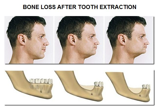
Tooronga Family Dentistry in Glen Iris

Family dental care in Glen Iris

Book Now

98227006
0450067475

Suite 1.02, 1 Crescent Rd., Glen Iris 3146
  • Home
  • About Us
    • Dr Daniel Kaufman – Experienced Dental Care Professional in Glen Iris
    • Why Visit Tooronga Family Dentistry in Glen Iris?
    • Frequently Asked Questions – Dentist Glen Iris
    • Our Practice in Glen Iris
    • State of the art materials and equipment in Glen Iris
    • Strict Infection Control at Tooronga Family Dentistry in Glen Iris
  • Services
    • Orthodontic braces
    • Straight Teeth with Clear aligners in Glen Iris
      • Invisalign Clear Aligners: A Discreet Alternative to Braces in Glen Iris
    • Wisdom tooth extraction
    • Fixing broken teeth.
    • Kids Dentist in Glen Iris
    • Healing bleeding Gums and bad breath
    • Mending teeth with a large cavities.
    • Teeth Whitening
    • Dealing with dental fear
    • Eliminating bad breath – Halitosis
    • Replacing missing teeth
    • Reinforcing fragile teeth in Glen Iris
    • Treating Molar and Incisor Hypomineralization
  • Contact
    • Emergency Contact
    • Book an Appointment Online
    • Ask Dr Kaufman a question
  • Patient information
    • Our Practice in Glen Iris
    • Blog
    • What is a Root canal treatment
    • Why remove decay from teeth
    • Oral hygiene in Glen Iris
    • Gum Disease During Pregnancy in Glen Iris: Causes, Symptoms, and Risks
    • Crowns and Bridges in Glen Iris
    • What to do for traumatic tooth injuries in Glen Iris
    • Tooth removal – Extractions
    • Child Dental Benefits Schedule
    • Professionally made mouthguard
    • Practice policies in Glen Iris
    • Privacy Policy
    • Terms and Conditions
    • Sitemap

TV-Featured Smile Makeovers: Porcelain Veneers in Glen Iris, Transform Your Smile

Posted on 02.16.26

Dream of a perfect smile like the stunning transformations featured on popular TV shows? The same smile makeover technology showcased on programs like The Doctors is now available right here in Glen Iris, helping local Melbourne residents achieve dramatic, life-changing results.

Porcelain veneers—the thin, custom-crafted facings that create Hollywood-worthy smiles—offer a versatile solution for multiple cosmetic dental concerns. Whether you’re dealing with gaps, discoloration, misshapen teeth, or worn edges, veneers can deliver the beautiful, confident smile you’ve always wanted without traveling to Melbourne’s CBD.

What Are Porcelain Veneers?

Porcelain veneers are ultra-thin shells of medical-grade ceramic that are permanently bonded to the front surface of your teeth. Think of them as custom-designed facings that completely transform your smile’s appearance while preserving most of your natural tooth structure.

The Science Behind the Beauty

Modern dental porcelain mimics the light-reflecting properties of natural tooth enamel with remarkable precision. This means your veneers won’t just look white—they’ll look naturally luminous, with the subtle translucency and depth that characterize a genuine, healthy smile.

Key Characteristics:

  • Thickness: Typically 0.5mm to 0.7mm (about as thin as a contact lens)
  • Material: High-quality dental porcelain or ceramic
  • Durability: Highly resistant to staining and chipping
  • Lifespan: 10-20 years or more with proper care
  • Appearance: Indistinguishable from natural teeth

Smile Transformations: What Veneers Can Achieve

The versatility of porcelain veneers makes them an ideal solution for numerous cosmetic concerns affecting Glen Iris patients:

1. Close Gaps and Spaces

The Problem: Gaps between teeth (diastema) can occur naturally or develop over time due to tooth movement, gum disease, or tooth loss. While some people embrace their gaps, others feel self-conscious about spaces in their smile.

The Veneer Solution: Veneers can close gaps without orthodontic treatment by:

  • Extending the width of teeth on either side of the space
  • Creating the appearance of perfectly aligned teeth
  • Eliminating spaces in a single treatment process
  • Providing immediate, permanent results

Treatment Time: 2-3 appointments over 2-3 weeks at our convenient Glen Iris location

2. Change Tooth Shape

The Problem: Misshapen teeth can result from:

  • Genetic factors affecting tooth development
  • Wear from grinding or clenching
  • Chips or fractures from injury
  • Uneven tooth lengths creating an irregular smile line
  • Teeth that appear too short, too long, too narrow, or too square

The Veneer Solution: Custom-designed veneers can reshape teeth by:

  • Creating ideal proportions between width and length
  • Establishing a harmonious, balanced smile line
  • Rounding sharp corners or squaring rounded edges
  • Restoring symmetry to your smile
  • Disguising minor rotations or tilting without orthodontics

Result: Teeth that appear naturally shaped and beautifully proportioned

3. Transform Tooth Color

The Problem: Tooth discoloration frustrates many people seeking a brighter smile. Common causes include:

  • Intrinsic staining from medications (like tetracycline), excessive fluoride, or tooth trauma
  • Age-related yellowing as enamel thins and underlying dentin shows through
  • Surface stains from coffee, tea, red wine, or tobacco that resist whitening treatments
  • Uneven coloring with spots, bands, or patches

The Veneer Solution: Porcelain veneers provide permanent color transformation that:

  • Whitening treatments cannot achieve for intrinsic stains
  • Covers discoloration completely and permanently
  • Resists future staining from foods, beverages, and lifestyle factors
  • Provides uniform, consistent color across all treated teeth
  • Can be customized to your preferred shade within natural-looking parameters

Important Note: Unlike natural teeth, veneers don’t respond to whitening treatments, so choose your desired shade thoughtfully during the design phase at our Glen Iris practice.

4. Lengthen Worn or Short Teeth

The Problem: Teeth can appear too short due to:

  • Excessive wear from grinding (bruxism)
  • Genetic factors resulting in naturally small teeth
  • Gum tissue covering too much of the tooth (gummy smile)
  • Age-related wear shortening teeth over time

Short teeth can make you appear older and may affect bite function.

The Veneer Solution: Veneers can lengthen teeth by:

  • Adding length to worn edges without damaging healthy tooth structure
  • Creating ideal tooth proportions for a more youthful appearance
  • Establishing proper vertical dimension for improved bite function
  • Balancing the ratio between visible tooth and gum tissue

Aesthetic Impact: Longer teeth create a more youthful, vigorous appearance and can even make your face look fuller and less aged.

5. Comprehensive Smile Makeovers

The Power of Multiple Veneers: While veneers can address individual tooth concerns, their most dramatic impact comes from comprehensive smile makeovers treating multiple teeth simultaneously.

Complete Transformation Possibilities:

  • Correct multiple concerns (color, shape, alignment, gaps) in one treatment plan
  • Create a unified, harmonious smile design
  • Achieve symmetry and ideal proportions across all visible teeth
  • Transform your entire appearance and boost confidence dramatically

Typical Coverage: Most smile makeovers involve 6-10 veneers on upper front teeth, though some Glen Iris patients choose to treat both upper and lower teeth for maximum impact.

The Glen Iris Veneer Process: Your Journey to a New Smile

Initial Consultation: Design Your Dream Smile

Your veneer journey begins with a comprehensive consultation at our Glen Iris dental practice, conveniently located in Melbourne’s eastern suburbs:

What Happens:

  1. Smile Analysis: We examine your teeth, gums, bite, and facial proportions
  2. Goal Discussion: You share your vision and concerns about your current smile
  3. Digital Imaging: Advanced technology shows potential results before treatment begins
  4. Custom Design: Together, we design your ideal smile considering your facial features, preferences, and lifestyle
  5. Treatment Planning: We outline the entire process, timeline, and investment required

Why This Matters: Veneers are a permanent cosmetic investment. Taking time to design your perfect smile ensures results you’ll love for years to come.

Convenient Location: Our Glen Iris practice is easily accessible from surrounding Melbourne suburbs including Ashburton, Camberwell, Malvern, Toorak, and Hawthorn, with convenient parking available.

Preparation Appointment: Creating Your Custom Veneers

The Process:

  1. Tooth Preparation: A minimal amount of enamel (typically 0.5mm) is removed to accommodate veneer thickness
  2. Precision Impressions: Detailed molds capture exact tooth dimensions and relationships
  3. Shade Selection: We carefully select porcelain color matching your desired outcome
  4. Temporary Veneers: Custom temporaries protect prepared teeth and preview your new smile
  5. Laboratory Fabrication: Expert Victorian ceramists handcraft your permanent veneers (typically 2-3 weeks)

Comfort Considerations:

  • Local anesthesia ensures a pain-free experience
  • Temporary veneers allow normal eating and speaking
  • Minimal tooth reduction preserves natural tooth strength

Bonding Appointment: Revealing Your New Smile

The Final Step:

  1. Veneer Trial: We place veneers without bonding to verify perfect fit, shape, and color
  2. Final Adjustments: Minor modifications ensure absolute precision
  3. Tooth Preparation: Teeth are cleaned and treated for optimal bonding
  4. Permanent Bonding: Each veneer is individually cemented using advanced adhesive technology
  5. Final Polishing: Edges are smoothed and polished for a natural, comfortable feel
  6. Bite Verification: We ensure proper alignment and comfortable function

The Moment of Truth: When you see your completed smile for the first time, the transformation is often emotionally overwhelming. Glen Iris patients frequently report feeling like they’re seeing their “real self” for the first time.

Why Glen Iris Patients Choose Veneers

Immediate, Dramatic Results

Unlike orthodontics requiring months or years, veneers deliver comprehensive smile transformation in just weeks. This makes them ideal for:

  • Upcoming weddings, reunions, or special events
  • Career advancement requiring polished professional appearance
  • Anyone tired of waiting for gradual improvement
  • People seeking maximum impact in minimum time

Conservative Tooth Preservation

Modern veneer preparation is remarkably conservative:

  • Minimal enamel removal (unlike crowns requiring extensive tooth reduction)
  • Tooth structure remains largely intact
  • Natural tooth strength is preserved
  • Most tooth sensitivity resolves quickly after placement

Long-Lasting Beauty

With proper care, quality porcelain veneers provide:

  • 10-20+ years of beautiful service
  • Exceptional stain resistance (won’t yellow like natural teeth)
  • Superior durability against chips and cracks
  • Color stability that maintains its beauty indefinitely

Natural-Looking Results

Today’s veneers are virtually indistinguishable from natural teeth due to:

  • Advanced porcelain formulations mimicking enamel translucency
  • Custom shading and characterization for realistic appearance
  • Attention to subtle details like texture and light reflection
  • Individualized design respecting your unique facial features

Versatility and Customization

Every veneer treatment is completely personalized:

  • No two smile makeovers are identical
  • Design reflects your personality, age, gender, and preferences
  • Can range from subtle enhancement to dramatic transformation
  • Achieves results impossible through other cosmetic methods

Caring for Your Veneers: Protecting Your Investment

Porcelain veneers are durable, but proper care ensures maximum longevity:

Daily Maintenance

Essential Habits:

  • Brush twice daily with non-abrasive toothpaste
  • Floss once daily using proper technique
  • Rinse after consuming staining foods or beverages
  • Avoid using teeth as tools (opening packages, biting nails, etc.)

Protecting Against Damage

Prevention Strategies:

  • Wear a night guard if you grind or clench teeth
  • Use a mouthguard during contact sports
  • Avoid biting extremely hard foods (ice, hard candy, bones)
  • Don’t use front teeth to tear or rip items

Professional Care

Regular Appointments at Our Glen Iris Practice:

  • Professional cleanings every six months
  • Periodic examinations to verify veneer integrity
  • Prompt attention to any concerns (loose veneers, chips, sensitivity)

What to Avoid

Habits That Damage Veneers:

  • Biting hard objects habitually
  • Using abrasive toothpastes or whitening products excessively
  • Neglecting oral hygiene leading to gum disease
  • Ignoring grinding issues that can fracture veneers

Are You a Candidate for Veneers?

Porcelain veneers work beautifully for most people, but ideal candidates typically have:

Good Overall Oral Health

  • Healthy gums free from active disease
  • No untreated tooth decay
  • Sufficient tooth enamel for bonding
  • Stable bite and jaw relationships

Specific Cosmetic Concerns

  • Discolored teeth resistant to whitening
  • Chipped, cracked, or worn teeth
  • Minor alignment or spacing issues
  • Disproportionate or misshapen teeth

Realistic Expectations

  • Understanding that veneers are permanent (enamel removal can’t be reversed)
  • Commitment to proper care and maintenance
  • Recognition that veneers may eventually need replacement
  • Appreciation for natural-looking rather than artificially perfect results

Situations Where Alternative Treatments May Be Better

Veneers might not be the best choice if you:

  • Have severe tooth misalignment (orthodontics may be preferable)
  • Grind teeth severely without willingness to wear a night guard
  • Have insufficient enamel from previous procedures
  • Need extensive structural tooth repair (crowns may be better)
  • Prefer completely reversible cosmetic options

During your Glen Iris consultation, we’ll honestly assess whether veneers are your best option or if alternative treatments might better serve your needs.

The Confidence Transformation: Beyond Just Teeth

The impact of a smile makeover extends far beyond aesthetics. Glen Iris patients who’ve completed veneer treatment consistently report:

Professional Benefits

  • Increased confidence in business settings and presentations across Melbourne
  • More positive first impressions with clients and colleagues
  • Enhanced professional image supporting career advancement
  • Greater comfort in video calls and professional photography

Social Advantages

  • Willingness to smile freely without self-consciousness at Glen Iris cafés and social venues
  • Improved confidence in dating and social situations
  • More natural, genuine expressions without hiding teeth
  • Enhanced appearance in photos and social media

Personal Impact

  • Elevated self-esteem and positive self-image
  • Reduced anxiety about dental appearance
  • Feeling younger and more vibrant
  • Alignment between inner self-perception and outer appearance

Emotional Wellbeing

  • Relief from years of smile-related insecurity
  • Pride in appearance rather than embarrassment
  • Increased willingness to engage socially
  • Overall improved quality of life

Investment in Your Smile

Porcelain veneers represent a significant investment in your appearance and confidence. During your Glen Iris consultation, we’ll provide:

  • Detailed cost breakdown for your specific treatment plan
  • Information about payment options and financing
  • Honest discussion of value relative to longevity and impact
  • Comparison with alternative treatments if applicable

Remember: While cost is a consideration, veneers lasting 15-20 years translate to just pennies per day for a transformation that impacts every aspect of your life.

Why Choose Our Glen Iris, Victoria Practice for Your Smile Makeover

Local Expertise and Experience

  • Serving the Glen Iris and eastern Melbourne communities for years
  • Specialized training in cosmetic dentistry
  • Extensive experience creating natural, beautiful veneer results for Victorian patients
  • Commitment to staying current with latest techniques and materials
  • Portfolio of successful smile transformations for Glen Iris residents

Advanced Technology

  • Digital smile design for previewing results
  • Precision impression materials ensuring perfect fit
  • Collaboration with master Victorian ceramists for superior aesthetics
  • State-of-the-art bonding protocols for lasting results

Personalized Approach

  • Individual attention to your unique goals and concerns
  • Custom smile design respecting your facial features
  • Patient education ensuring informed decisions
  • Ongoing support throughout treatment and beyond

Comprehensive Care

  • Treatment of any underlying oral health issues first
  • Holistic approach considering function and aesthetics
  • Coordination with specialists when needed
  • Long-term maintenance planning

Convenient Eastern Melbourne Location

Easy Access from Throughout Melbourne’s East: Our Glen Iris dental practice is conveniently located for patients from:

  • Glen Iris and surrounding suburbs
  • Ashburton, Camberwell, Canterbury
  • Malvern, Toorak, Armadale
  • Hawthorn, Kew, Kooyong
  • Burwood, Chadstone, Mount Waverley
  • And throughout Melbourne’s eastern suburbs

Accessibility Features:

  • Convenient parking available
  • Public transport access via Gardiner Station and local tram routes
  • Flexible appointment scheduling to accommodate work and family commitments
  • Evening and Saturday appointments available

Ready to Transform Your Smile in Glen Iris?

The beautiful, confident smile you’ve seen on television and admired in others is within your reach right here in Glen Iris. Porcelain veneers have helped thousands of Melbourne residents achieve smile transformations that changed their lives—and the same technology is available at our convenient eastern suburbs location.

Whether you’re dealing with discolored teeth that won’t whiten, gaps you’d like to close, misshapen teeth, or simply want a more attractive smile, veneers may be your ideal solution.

The first step is discovering what’s possible for your unique smile—without traveling to Melbourne’s CBD.


Our Glen Iris, Victoria dental practice is dedicated to creating smile transformations that look natural, feel comfortable, and last for years. We combine artistic vision with clinical excellence to deliver results that exceed expectations while respecting your individual features and preferences. As a trusted part of the Glen Iris community, we’re committed to providing world-class cosmetic dentistry close to home.

Call or book online Tooronga Family Dentistry on (03) 9822 7006 to Schedule Your Smile Makeover Consultation Today – Contact our Glen Iris dental clinic to begin your journey toward the confident, beautiful smile you deserve. During your consultation at our convenient eastern Melbourne location, we’ll discuss your goals, show you what’s possible, and design a treatment plan tailored specifically for you.

See What Veneers Can Do for Your Smile – Don’t spend another day feeling self-conscious about your teeth. Call our Glen Iris, Victoria practice now or book your consultation online to discover how porcelain veneers can transform your appearance and your life—all without leaving Melbourne’s eastern suburbs.

Teeth Grinding from Stress in Glen Iris: How Clenching Destroys Teeth and Why You Need a Night Guard

Posted on 02.10.26

Understanding Teeth Grinding and Stress

Teeth grinding stress is destroying your teeth right now—whether you know it or not. Stress is an unfortunate part of our life, one of its manifestations is tooth clenching and grinding. At Tooronga Family Dentistry, Dr. Kaufman sees the devastating effects of stress-induced teeth grinding daily: cracked teeth, worn enamel, broken fillings, and even tooth loss. Understanding how stress causes teeth grinding, why teeth grinding damages enamel permanently, and how night guards prevent teeth grinding destruction empowers Glen Iris patients to protect their smiles before irreversible damage occurs.


Why Teeth Grinding Happens: The Stress Connection

Stress Causes Teeth Grinding

Stress is the leading cause of teeth grinding (bruxism). When you experience stress, your body responds with muscle tension—including your jaw muscles. This muscle tension from stress causes teeth clenching and teeth grinding, often without your awareness.

Stress teeth grinding statistics:

  • 70% of teeth grinding cases caused by stress and anxiety
  • 80% of teeth grinding occurs during sleep (unconscious)
  • 30-40 million Americans grind teeth (many due to stress)
  • Women grind teeth from stress more than men (ratio 2:1)

How Your Mouth Is Designed: Teeth Should Not Touch

Our mouth has evolved in such a way that it stays slightly ajar and the teeth don’t touch usually.

Normal teeth position:

✓ Teeth slightly apart (1-3mm gap—natural resting position) ✓ Tongue on palate (gentle contact—no tooth pressure) ✓ Lips closed (or slightly parted—relaxed jaw) ✓ No muscle tension (jaw muscles relaxed—energy conserved)

Why teeth staying apart protects enamel:

This way the teeth can retain the strong enamel cap for longer and it is not worn down.

When teeth don’t touch except during chewing/swallowing:

  • Enamel preserved (no friction—surface intact)
  • No pressure stress (enamel not compressed—prevents microcracks)
  • Longer tooth lifespan (decades of function—minimal wear)

When Teeth Should Touch

The only time teeth are meant to come together is when chewing or swallowing.

Normal tooth contact:

✓ Chewing: 15-20 minutes daily (meals—brief, intermittent contact) ✓ Swallowing: 600-1000 times daily (each contact <1 second—minimal total time) ✓ Total daily contact: 20-30 minutes maximum (healthy, designed-for contact)

Teeth grinding comparison:

⚠ Grinding/clenching: 20-40 minutes to several hours nightly ⚠ Force: 400-800 PSI (vs. 150-200 PSI chewing—excessive, destructive) ⚠ Total contact time: 10-20x normal (overwhelming designed capacity)

The problem: Teeth grinding from stress creates excessive tooth contact—far beyond evolutionary design, causing permanent enamel damage.


What Happens During Teeth Grinding and Clenching

Clenching: Stress Muscle Contraction

When we are stressed, among other things our main chewing muscles contract and the teeth meet and press against each other, this is clenching.

The stress clenching mechanism:

  1. Stress response (fight-or-flight activation—cortisol, adrenaline release)
  2. Muscle tension (throughout body—including jaw)
  3. Masseter muscle contraction (main chewing muscle—clamping teeth together)
  4. Temporalis muscle contraction (temple muscle—reinforcing clench)
  5. Sustained pressure (teeth pressed together—400+ PSI force)

Clenching vs. grinding:

  • Clenching: Teeth pressed together (static—held forcefully)
  • Grinding: Teeth sliding (dynamic—lateral/forward motion)
  • Often both: Clenching plus grinding (combined destruction)

Grinding (Bruxism): The Lateral Motion

In some of us in addition to clenching other muscles start to pull the lower jaw forward and sideways leading to the grinding motion or bruxism.

The grinding mechanism:

Additional muscles activating:

  • Lateral pterygoid (pulling jaw forward—protrusive motion)
  • Medial pterygoid (pulling jaw sideways—lateral motion)
  • Combined action: Jaw moving in circular or lateral grinding pattern

Grinding motion types:

⚠ Side-to-side grinding (most common—teeth sliding laterally) ⚠ Forward-backward grinding (protrusive—teeth sliding front/back) ⚠ Circular grinding (combination—most destructive)

Why grinding worse than clenching:

  • Clenching: Vertical pressure only (compression—enamel strong in this direction)
  • Grinding: Lateral shearing forces (enamel weak against—accelerated wear)
  • Grinding damage: 10x faster enamel loss vs. clenching alone

Lifestyle Factors Triggering Teeth Grinding

This process can be triggered by other lifestyle factors like smoking, large caffeine intake and heavy alcohol consumption or by taking antidepressants.

Teeth grinding triggers beyond stress:

1. Smoking

  • Nicotine stimulant (muscle activation—increased grinding)
  • Dopamine disruption (affecting motor control—bruxism intensity)
  • Smokers 2x more likely to grind teeth vs. non-smokers

2. Large Caffeine Intake

  • Stimulant effect (muscle activity—jaw tension)
  • Sleep disruption (poor sleep quality—increased night grinding)
  • >4 cups daily: Significant teeth grinding risk increase

3. Heavy Alcohol Consumption

  • Muscle relaxation (during drinking—jaw dropping)
  • Rebound activation (during sleep—muscles overcompensating, grinding)
  • Sleep disruption (REM interference—grinding during sleep transitions)

4. Antidepressants

  • SSRIs particularly (Prozac, Zoloft, Paxil—common bruxism side effect)
  • Serotonin effect (neurotransmitter—affecting motor control)
  • 10-20% of SSRI users experience teeth grinding
  • Dose-dependent: Higher doses = more grinding

Combined effects: Stress + smoking + caffeine + alcohol + antidepressants = severe teeth grinding (multiple triggers compounding)


The Destruction: How Teeth Grinding Damages Teeth Permanently

Stage 1: Enamel Microcracks from Clenching

The clenching leads to the formation of minute cracks in the enamel:

Microcrack formation:

  1. Excessive force (clenching 400-800 PSI—overwhelming enamel)
  2. Enamel compression (crystalline structure stressed—microscopic fractures)
  3. Microcracks form (invisible initially—within enamel layer)
  4. Stress concentration (cracks becoming weak points—propagating further)

Microcrack characteristics:

  • Microscopic initially (not visible—only detectable with magnification/transillumination)
  • Progressive (each clench cycle—expanding slightly)
  • Irreversible (enamel cannot heal—unlike bone, skin)

Stage 2: Visible Cracks Developing

Which over time develop to larger visible cracks:

Crack progression timeline:

Months 1-6: Microcracks accumulating (undetectable—patient unaware) Months 6-18: Cracks coalescing (occasionally visible—fine lines on enamel) Years 2-5: Large visible cracks (obvious—running across tooth surface) Years 5+: Through-and-through cracks (extending to dentin, pulp—tooth fracture risk)

Visible crack appearance:

  • Craze lines (surface cracks—enamel only, often stained)
  • Fractured cusp lines (deeper cracks—destabilizing tooth portions)
  • Vertical cracks (most serious—extending toward root)

Stage 3: Cracks Extending to Dentin

And extend to the dentine which is the inner layer of the tooth:

Why dentin involvement serious:

⚠ Dentin softer (than enamel—cracks propagating faster once reached) ⚠ Tubules present (microscopic channels—bacteria accessing, sensitivity) ⚠ Near pulp (nerve chamber—infection risk, pain) ⚠ Structural weakness (crack compromising—tooth fracture imminent)

Dentin crack consequences:

  • Sensitivity (cold, hot, sweet—stimuli reaching nerve through tubules)
  • Decay risk (bacteria entering cracks—cavity formation)
  • Fracture vulnerability (weakened structure—cusp breaking off)
  • Root canal need (if crack reaches pulp—infection, tooth death)

Stage 4: Tooth Chipping and Wear from Grinding

The grinding or bruxing of the teeth leads to tooth chipping, wear and loss of tooth.

Grinding damage types:

Chipping

⚠ Cusp fractures (pointed portions breaking—from lateral grinding forces) ⚠ Edge fractures (front teeth edges—chipping, shortening) ⚠ Filling fractures (restorations breaking loose—concentrated stress)

Wear (Attrition)

⚠ Flattened cusps (molars ground flat—losing chewing surface anatomy) ⚠ Shortened teeth (enamel worn away—teeth appearing shorter) ⚠ Smooth, shiny surfaces (natural texture polished away—”ground down” appearance) ⚠ Dentin exposure (enamel completely worn through—yellow inner layer visible)

Tooth Loss

⚠ Fracture beyond repair (crack extending to root—extraction needed) ⚠ Severe wear (tooth worn to gum line—non-restorable) ⚠ Multiple teeth (years of grinding—extensive tooth loss)


The Permanent Nature of Teeth Grinding Damage

Once the teeth have cracked or have been lost they will not heal back like broken skin or broken bone and they will continue to deteriorate.

Why teeth don’t heal:

Bone/skin healing: ✓ Living tissue (blood supply—delivering repair cells) ✓ Regeneration capacity (new tissue forming—filling fracture) ✓ Complete healing (weeks to months—restored strength)

Tooth structure: ✗ Enamel acellular (no living cells—cannot regenerate) ✗ Dentin minimal repair (limited, slow—inadequate for cracks) ✗ No blood supply (enamel—no healing mechanism) ✗ Permanent damage (cracks, wear never reversing—only worsening)

The progressive deterioration:

  1. Crack forms (from grinding/clenching)
  2. Crack propagates (each grinding episode—extending further)
  3. Eventual fracture (crack reaching critical length—tooth breaking)
  4. No healing (damage accumulating—irreversible)

The reality: Every night of teeth grinding causes permanent damage—accumulating over time until tooth requires extraction.


The Solution: Night Guards Prevent Teeth Grinding Destruction

How Night Guards Work

To prevent the destruction we need to separate the teeth when they are clenched and guide them in non-destructive paths when the muscles pull the jaw sideways.

Night guard protective mechanisms:

Function 1: Separating Teeth During Clenching

This is achieved with an occlusal splint or a night guard.

✓ Physical barrier (between upper/lower teeth—preventing direct contact) ✓ Force absorption (night guard material—compressing instead of enamel) ✓ Even distribution (spreading clenching force—across all teeth, not concentrated) ✓ Enamel preservation (no tooth-on-tooth contact—zero enamel wear)

Separation benefits:

  • No microcracks (teeth not touching—no compression damage)
  • No wear (grinding on plastic—not enamel)
  • Existing damage halted (cracks not propagating—deterioration stopped)

Function 2: Guiding Jaw in Non-Destructive Paths

Guide them in non-destructive paths when the muscles pull the jaw sideways:

✓ Smooth surface (allowing lateral slide—no catching, shearing) ✓ Controlled movement (designed pathways—reducing destructive forces) ✓ Reduced muscle activity (some evidence night guards—decreasing grinding intensity)

Guidance benefits:

  • Lateral forces minimized (smooth gliding—not rough grinding)
  • Even wear on guard (plastic wearing—not teeth)
  • Muscle “reprogramming” (over time—reduced grinding activity)

Night Guard Types

Occlusal splint or night guard options:

Custom Hard Night Guards (Dr. Kaufman Provides)

✓ Hard acrylic (rigid—superior force distribution) ✓ Custom fit (precise—comfortable, stays in place) ✓ Thin design (2-3mm—comfortable wear) ✓ Durable (3-5+ years—long-term protection) ✓ Proven effective (research-validated—preventing tooth damage)

Cost: $400-600 (preventing $thousands in tooth repair)

Over-the-Counter Soft Night Guards

⚠ Soft material (compresses—limited protection) ⚠ Poor fit (boil-and-bite—uncomfortable, falls out) ⚠ Thick (4mm+—gag reflex, discomfort) ⚠ Short lifespan (3-6 months—frequent replacement) ⚠ May increase grinding (chewable texture—stimulating jaw activity)

The verdict: Custom hard night guards dramatically superior for teeth grinding protection.


Teeth Grinding Symptoms: Do You Grind?

Signs You’re Grinding Your Teeth from Stress

Teeth grinding symptoms:

🦷 Morning jaw pain (sore muscles—from night grinding) 🦷 Headaches (temples, neck—muscle tension) 🦷 Worn, flattened teeth (cusps ground down—visible damage) 🦷 Tooth sensitivity (cold, hot—enamel thinned from wear) 🦷 Cracked teeth (visible lines—grinding damage) 🦷 Chipped teeth (edges broken—grinding fractures) 🦷 Loose teeth (grinding forces—damaging periodontal ligament) 🦷 Partner reports grinding sounds (loud—audible grinding during sleep) 🦷 Tongue indentations (scalloped edges—pressed against teeth during clenching) 🦷 Cheek ridges (white lines inside cheek—chronic pressing against teeth)

If 3+ symptoms: Likely grinding—need night guard evaluation.


Preventing Teeth Grinding: Beyond Night Guards

Stress Management for Teeth Grinding

Addressing root cause:

✓ Stress reduction (meditation, therapy, exercise—reducing grinding trigger) ✓ Sleep hygiene (consistent schedule, cool room, no screens—improving sleep quality) ✓ Caffeine reduction (limiting to morning—avoiding afternoon/evening) ✓ Alcohol moderation (limiting intake—especially before bed) ✓ Smoking cessation (quitting—removing major grinding trigger)

Medication Adjustment

If antidepressants causing grinding:

  • Discuss with doctor (possibly adjusting dose, switching medications)
  • Timing change (morning instead of night—reducing sleep grinding)
  • Additional medication (buspirone sometimes prescribed—reducing SSRI bruxism)

Never stop medications without doctor supervision.

Jaw Exercises and Physical Therapy

Reducing muscle tension:

✓ Gentle stretching (opening wide, side-to-side—reducing tightness) ✓ Massage (masseter, temporalis—releasing muscle knots) ✓ Heat application (warm compress—relaxing muscles) ✓ Physical therapy (specialized TMJ therapy—if severe)


Expert Teeth Grinding Treatment in Glen Iris

Dr. Kaufman’s Comprehensive Bruxism Care

Tooronga Family Dentistry provides:

✓ Teeth grinding evaluation (examining wear, cracks, jaw—assessing damage) ✓ Custom night guard fabrication (precise fit—maximum protection) ✓ Stress counseling (identifying triggers—holistic approach) ✓ Damaged tooth repair (fillings, crowns—restoring fractured teeth) ✓ TMJ evaluation (jaw joint assessment—addressing pain) ✓ Follow-up monitoring (checking night guard—ensuring effectiveness)

Why choose Tooronga Family Dentistry for teeth grinding:

  • Evidence-based (custom hard night guards—proven superior)
  • Comprehensive (addressing causes—not just symptoms)
  • Experienced (Dr. Kaufman—treating teeth grinding daily)
  • Preventive focus (stopping damage—before extensive repair needed)
  • Glen Iris location (convenient—serving Malvern, Ashburton, Camberwell)

Schedule Your Teeth Grinding Consultation

Stop Teeth Grinding Damage Today

Protect your teeth from stress grinding destruction.

Call Tooronga Family Dentistry: 9822 7006

What to Expect

  1. Comprehensive examination (teeth, jaw, muscles—damage assessment)
  2. Grinding severity evaluation (wear patterns—determining treatment urgency)
  3. Stress discussion (lifestyle factors—identifying triggers)
  4. Custom night guard impressions (precise molds—ensuring perfect fit)
  5. Treatment plan (protecting teeth—preventing further damage)
  6. Follow-up scheduling (fitting appointment—ongoing monitoring)

Contact Information

  • Phone: 9822 7006
  • Services: Teeth grinding treatment, custom night guards, stress bruxism care
  • Location: Glen Iris, serving Malvern, Ashburton, Camberwell, surrounding Melbourne

Teeth Grinding FAQs

Can teeth grinding be cured?

Teeth grinding often can’t be “cured” (especially stress-related), but damage prevention with night guards is highly effective. Stress management may reduce grinding intensity.

Do night guards stop teeth grinding?

Night guards don’t stop grinding—they protect teeth from grinding damage. Grinding continues, but forces dissipate on guard instead of destroying enamel.

How much does teeth grinding damage cost to repair?

Without night guard: $5,000-15,000+ (multiple crowns, possible implants from tooth loss) With night guard: $400-600 initial + minimal repair (prevention dramatically cheaper)

Can stress teeth grinding cause tooth loss?

Yes. Severe stress teeth grinding causes cracks extending to roots—fracturing teeth beyond repair, requiring extraction.

How long do custom night guards last?

Custom hard night guards: 3-5+ years (some patients 10+ years) Soft OTC guards: 3-6 months (frequent replacement needed)


Take Action: Protect Your Teeth from Stress Grinding

The Bottom Line

Stress causes teeth grinding—and teeth grinding destroys teeth permanently:

  • ⚠ Microcracks form (enamel compressed—invisible damage starting)
  • ⚠ Cracks grow (each grinding night—propagating toward dentin)
  • ⚠ Teeth wear down (lateral grinding—enamel abrading)
  • ⚠ Teeth fracture (cracks reaching critical length—cusps breaking)
  • ⚠ Teeth lost (beyond repair—extraction needed)
  • ⚠ Damage permanent (teeth can’t heal—deterioration continuing)

Night guards prevent destruction:

  • ✅ Separate teeth (no direct contact—no compression microcracks)
  • ✅ Guide jaw (smooth lateral movement—no destructive grinding)
  • ✅ Protect enamel (wearing plastic—not teeth)
  • ✅ Stop deterioration (halting damage—before tooth loss)

Investment: $400-600 custom night guard prevents $10,000+ in future dental work.

Don’t wait until teeth crack, chip, or fracture.

Call 9822 7006 today.

Dr. Kaufman will evaluate your teeth grinding, fabricate custom night guard, and stop stress from destroying your teeth.

Serving Glen Iris, Malvern, Ashburton, Camberwell with expert teeth grinding treatment.

Your teeth can’t heal. Protect them now.

Gum Disease in Glen Iris: Link to Heart Disease, Diabetes, and Whole-Body Health

Posted on 02.4.26

Understanding Gum Disease: More Than Just Your Mouth

Gum disease affects far more than teeth—threatening heart health, increasing diabetes risk, and causing systemic inflammation throughout the body. At Tooronga Family Dentistry, Dr. Kaufman treats gum disease with urgency, understanding that several studies have shown that periodontal disease is associated with heart disease—making gum disease a serious medical condition, not just a dental problem. Scientists believe that the inflammation caused by periodontal disease may be responsible for the association between gum disease and heart disease, diabetes, and other systemic conditions. Understanding how gum disease affects the body, recognizing gum disease symptoms, knowing gum disease and heart disease connection, and getting regular gum disease screening empowers Glen Iris patients to protect both oral and overall health.


Gum Disease and Heart Disease: The Dangerous Connection

The Scientific Evidence Linking Gum Disease to Heart Disease

Several studies have shown that periodontal disease is associated with heart disease:

Gum disease-heart disease research:

📊 2-3x higher heart disease risk (people with gum disease—vs. healthy gums) 📊 American Heart Association (confirms gum disease association—independent risk factor) 📊 Meta-analysis of 59 studies (gum disease linked to cardiovascular events—heart attacks, strokes) 📊 Dose-response relationship (severe gum disease—higher heart disease risk than mild)

Types of heart disease linked to gum disease:

⚠ Coronary artery disease (atherosclerosis—gum disease bacteria in plaques) ⚠ Heart attack (myocardial infarction—gum disease increasing risk) ⚠ Stroke (ischemic—gum disease causing blood vessel blockages) ⚠ Atherosclerosis (arterial plaque buildup—gum disease contributing) ⚠ Endocarditis (heart valve infection—gum disease bacteria seeding)


How Gum Disease Causes Heart Disease: The Inflammation Pathway

Scientists believe that the inflammation caused by periodontal disease may be responsible for the association:

Gum disease inflammation mechanism:

Local gum disease inflammation:

⚠ Bacterial infection (gum disease bacteria in gum pockets—Porphyromonas gingivalis, Tannerella forsythia) ⚠ Immune response (white blood cells fighting—gum disease triggering) ⚠ Inflammatory mediators released: IL-1, IL-6, TNF-alpha, C-reactive protein (gum disease chemicals) ⚠ Chronic inflammation (ongoing gum disease—never resolving, constant inflammatory state)

Systemic inflammation from gum disease:

⚠ Bacteria entering bloodstream (bacteremia—gum disease bacteria through inflamed gums) ⚠ Inflammatory chemicals circulating (gum disease mediators—affecting entire body) ⚠ C-reactive protein elevated (blood marker—gum disease causing systemic inflammation) ⚠ Endothelial dysfunction (blood vessel lining damage—gum disease impairing)

Gum disease triggering atherosclerosis:

  1. Gum disease bacteria in bloodstream (bacteremia—daily with chewing, brushing)
  2. Bacteria adhere to blood vessel walls (gum disease colonizing arteries)
  3. Inflammation in vessel walls (gum disease immune response—damaging endothelium)
  4. Plaque formation (cholesterol, inflammatory cells—gum disease accelerating atherosclerosis)
  5. Narrowed arteries (restricted blood flow—gum disease contributing to heart attack, stroke)

Porphyromonas Gingivalis: The Gum Disease Bacteria Behind Heart Disease

The Specific Gum Disease Bacteria-Heart Connection

Research shows fascinating features of a bacteria, Porphyromonas gingivalis, who has been known to cause gum disease:

Porphyromonas gingivalis (P. gingivalis):

✓ Primary gum disease pathogen (keystone species—driving gum disease progression) ✓ Gram-negative anaerobic (thrives without oxygen—in deep gum disease pockets) ✓ Black-pigmented (produces dark pigment—visible in severe gum disease) ✓ Highly virulent (aggressive—gum disease destruction)

Prevalence in gum disease:

  • Found in 85-90% of severe gum disease cases (hallmark pathogen)
  • Absent in healthy gums (specific to gum disease—not normal flora)

How P. Gingivalis from Gum Disease Affects Blood Vessels

The bacteria activate the immune system and causes the cells to aggregate together and form blockages in the blood vessels:

P. gingivalis mechanisms in gum disease and heart disease:

Mechanism 1: Immune system activation by gum disease bacteria

The bacteria activate the immune system:

⚠ P. gingivalis entering bloodstream (from gum disease pockets—bacteremia) ⚠ White blood cells responding (neutrophils, macrophages—attacking gum disease bacteria) ⚠ Inflammatory cascade (cytokine storm—gum disease triggering IL-1β, IL-6, TNF-α) ⚠ Chronic immune activation (persistent gum disease—constant low-grade inflammation)

Mechanism 2: Cell aggregation and blockages from gum disease

And causes the cells to aggregate together and form blockages in the blood vessels:

⚠ Platelet aggregation (P. gingivalis—causing platelets to clump, gum disease clotting) ⚠ Foam cell formation (macrophages engulfing cholesterol—gum disease atherosclerotic plaques) ⚠ Thrombus formation (blood clots—gum disease bacteria triggering) ⚠ Arterial blockages (plaques, clots—gum disease causing heart attacks, strokes)

Specific P. gingivalis virulence factors in gum disease:

  • Gingipains (proteases—destroying tissue, activating coagulation from gum disease)
  • Fimbriae (adhesion structures—attaching to blood vessel walls, gum disease bacteria colonizing)
  • LPS (lipopolysaccharide) (endotoxin—triggering massive inflammation, gum disease immune response)

Research Findings: P. Gingivalis from Gum Disease in Atherosclerotic Plaques

Groundbreaking gum disease-heart disease studies:

📊 P. gingivalis DNA found in atherosclerotic plaques (60-70% of samples—gum disease bacteria in heart arteries) 📊 Live P. gingivalis cultured from plaques (not just dead bacteria—active gum disease infection in arteries) 📊 Higher P. gingivalis burden = more severe atherosclerosis (correlation—gum disease bacterial load) 📊 Animal studies: P. gingivalis infection (accelerating atherosclerosis—gum disease causation evidence)

How P. gingivalis from gum disease reaches arteries:

  1. Gum disease pocket bleeding (brushing, chewing, flossing—inflamed gums)
  2. Bacteremia (P. gingivalis entering bloodstream—from gum disease)
  3. Traveling through circulation (gum disease bacteria—to coronary, carotid arteries)
  4. Adhering to vessel walls (fimbriae—gum disease bacteria colonizing)
  5. Invading endothelial cells (P. gingivalis intracellular—gum disease bacteria hiding, persisting)
  6. Triggering plaque formation (inflammation, foam cells—gum disease atherosclerosis)

Gum Disease and Diabetes: The Two-Way Connection

How Gum Disease Initiates and Worsens Diabetes

This feature suggests that gum disease may initiate a heart disease in a similar manner that it can lead to diabetes:

Gum disease-diabetes bidirectional relationship:

Gum disease worsening diabetes:

⚠ Chronic inflammation from gum disease (IL-6, TNF-α—insulin resistance) ⚠ Elevated blood sugar (gum disease inflammation—impairing glucose control) ⚠ HbA1c increase (0.4-1.0%—gum disease worsening diabetes control) ⚠ Diabetic complications accelerated (gum disease—kidney disease, neuropathy faster)

Gum disease potentially triggering diabetes:

⚠ Systemic inflammation (gum disease—insulin resistance developing) ⚠ Beta cell dysfunction (pancreas—gum disease inflammation damaging) ⚠ Pre-diabetes → diabetes (gum disease pushing over threshold)

Evidence for gum disease-diabetes connection:

📊 Diabetics 2-3x more likely to have gum disease (vs. non-diabetics) 📊 Gum disease treatment improves HbA1c (0.4% reduction—gum disease therapy helping diabetes) 📊 Severe gum disease predicts diabetes development (longitudinal studies—gum disease preceding diagnosis)


The Parallel Pathways: Gum Disease to Heart Disease and Diabetes

Similar mechanisms:

Gum disease → Heart disease:

  1. P. gingivalis bacteremia (gum disease bacteria in blood)
  2. Inflammation in blood vessels (gum disease triggering)
  3. Platelet aggregation, plaque formation (gum disease causing blockages)
  4. Heart attack, stroke (gum disease outcome)

Gum disease → Diabetes:

  1. P. gingivalis chronic infection (gum disease ongoing)
  2. Systemic inflammation (gum disease cytokines)
  3. Insulin resistance (gum disease impairing glucose metabolism)
  4. Type 2 diabetes (gum disease contributing)

Common thread: Chronic inflammation from gum disease


Other Systemic Conditions Linked to Gum Disease

Gum Disease Affects the Whole Body

Additional health conditions associated with gum disease:

Respiratory diseases from gum disease:

⚠ Pneumonia (aspiration—gum disease bacteria into lungs) ⚠ COPD (chronic obstructive pulmonary disease—gum disease worsening) ⚠ Respiratory infections (gum disease bacteria reservoir)

Pregnancy complications from gum disease:

⚠ Preterm birth (2-7x higher risk—gum disease inflammation) ⚠ Low birth weight (gum disease affecting fetal development) ⚠ Pre-eclampsia (gum disease contributing)

Alzheimer’s disease and gum disease:

⚠ P. gingivalis in brain tissue (Alzheimer’s patients—gum disease bacteria crossing blood-brain barrier) ⚠ Gingipains in neurons (gum disease toxins—neurodegeneration) ⚠ 2x higher Alzheimer’s risk (with gum disease—longitudinal studies)

Rheumatoid arthritis and gum disease:

⚠ Shared inflammatory pathways (gum disease and RA—IL-1, TNF-α) ⚠ P. gingivalis producing citrullinated proteins (RA autoantigen—gum disease triggering RA) ⚠ Bidirectional worsening (gum disease worsening RA, RA worsening gum disease)

Kidney disease from gum disease:

⚠ Chronic kidney disease risk (increased with gum disease—inflammation damaging) ⚠ End-stage renal disease (higher with severe gum disease)


Gum Disease Symptoms: Early Detection

Recognizing Gum Disease Before It’s Too Late

For these reasons it is important to have your gums regularly checked to detect any signs of inflammation:

Early gum disease symptoms (gingivitis):

⚠ Bleeding gums (brushing, flossing—gum disease inflammation) ⚠ Red, swollen gums (vs. healthy pink—gum disease acute) ⚠ Tender gums (sensitive to touch—gum disease beginning) ⚠ Bad breath (halitosis—gum disease bacteria producing)

Moderate gum disease symptoms (early periodontitis):

⚠ Gum recession (teeth appearing longer—gum disease bone loss) ⚠ Pockets forming (>4mm—gum disease detachment) ⚠ Increased sensitivity (exposed roots—from gum disease) ⚠ Spontaneous bleeding (without brushing—gum disease severe inflammation)

Advanced gum disease symptoms (severe periodontitis):

⚠ Loose teeth (mobility—gum disease bone destruction) ⚠ Shifting teeth (drifting, spacing—gum disease support loss) ⚠ Pus between teeth and gums (abscess—gum disease infection) ⚠ Pain chewing (tooth movement—advanced gum disease) ⚠ Tooth loss (falling out—end-stage gum disease)

Silent gum disease:

⚠ Often painless (gum disease progressing—no symptoms until advanced) ⚠ Smokers especially (reduced bleeding—masking gum disease signs) ⚠ Regular checkups essential (detecting gum disease early—before symptoms)


Gum Disease Screening and Diagnosis in Glen Iris

Comprehensive Gum Disease Evaluation

Dr. Kaufman’s gum disease screening:

Visual examination for gum disease:

✓ Gum color (healthy pink vs. red—gum disease inflammation) ✓ Gum contour (knife-edge vs. swollen—gum disease edema) ✓ Bleeding (probing—gum disease sign) ✓ Recession (measuring—gum disease attachment loss)

Periodontal probing for gum disease:

✓ Measuring pocket depths (around every tooth—gum disease severity) ✓ Healthy: 1-3mm (no gum disease) ✓ Gingivitis: 3-4mm (early gum disease, reversible) ✓ Mild periodontitis: 4-5mm (established gum disease, bone loss beginning) ✓ Moderate periodontitis: 5-6mm (progressive gum disease) ✓ Severe periodontitis: 7+mm (advanced gum disease, significant bone loss)

X-rays for gum disease:

✓ Bone level assessment (radiographs—gum disease destruction visible) ✓ Bone loss patterns (horizontal, vertical—gum disease severity) ✓ Furcation involvement (molars—gum disease between roots)

Risk factor assessment for gum disease:

✓ Smoking (4-6x higher gum disease risk) ✓ Diabetes (2-3x higher gum disease risk, worse outcomes) ✓ Family history (genetic susceptibility—gum disease risk) ✓ Medications (dry mouth—increasing gum disease) ✓ Stress (immune suppression—worsening gum disease)


Gum Disease Treatment: Stopping the Inflammation

Treating Gum Disease to Protect Oral and Systemic Health

Gum disease treatment stages:


Stage 1: Professional Gum Disease Cleaning

For gingivitis and early gum disease:

✓ Scaling (removing plaque, tartar—gum disease bacterial deposits) ✓ Root planing (smoothing roots—gum disease toxin removal) ✓ Polishing (removing stains—gum disease prevention) ✓ Fluoride (strengthening enamel—gum disease sensitivity)

Frequency for gum disease:

  • Gingivitis: Every 6 months (preventing gum disease progression)
  • Controlled periodontitis: Every 3-4 months (maintaining gum disease remission)

Stage 2: Deep Cleaning for Gum Disease (Scaling and Root Planing)

For moderate to severe gum disease:

✓ Quadrant scaling/root planing (deep cleaning—gum disease pockets >4mm) ✓ Local anesthesia (numbing—comfortable gum disease treatment) ✓ Removing subgingival calculus (below gumline—gum disease deposits) ✓ Bacterial reduction (85-90%—gum disease infection controlled)

Gum disease healing after treatment:

  • Pocket reduction: 1-2mm (4mm → 2-3mm—gum disease improving)
  • Bleeding reduction: 80-90% (inflammation resolving—gum disease healing)
  • Timeframe: 4-6 weeks (tissue healing—gum disease response)

Stage 3: Advanced Gum Disease Treatment

For severe or refractory gum disease:

✓ Antibiotics (systemic or local—gum disease bacterial suppression)

  • Doxycycline (pills or gel—anti-P. gingivalis, gum disease treatment)
  • Amoxicillin + metronidazole (combination—severe gum disease) ✓ Gum surgery (flap surgery—gum disease deep pocket access) ✓ Bone grafting (regenerating bone—gum disease destruction repair) ✓ Guided tissue regeneration (membranes—gum disease lost attachment regrowth)

Stage 4: Gum Disease Maintenance

Lifelong gum disease management:

✓ Periodontal maintenance cleanings (every 3 months—gum disease control) ✓ Pocket monitoring (measuring depths—gum disease recurrence detection) ✓ Home care optimization (brushing, flossing, interdental brushes—gum disease prevention) ✓ Risk factor modification (smoking cessation, diabetes control—gum disease reduction)


Preventing Gum Disease: Daily Habits

Protecting Your Gums and Your Heart

Home care for gum disease prevention:

✓ Brushing twice daily (2 minutes, soft bristles—removing gum disease plaque) ✓ Flossing daily (interdental cleaning—gum disease prevention between teeth) ✓ Interdental brushes (for spaces—gum disease hard-to-reach areas) ✓ Antimicrobial mouthwash (chlorhexidine, essential oils—gum disease bacterial reduction) ✓ Electric toothbrush (superior plaque removal—gum disease prevention)

Lifestyle modifications for gum disease:

✓ Quit smoking (single biggest factor—gum disease risk reduction 50-80%) ✓ Control diabetes (blood sugar management—gum disease improvement) ✓ Stress reduction (immune support—gum disease resistance) ✓ Healthy diet (anti-inflammatory—omega-3s, antioxidants reducing gum disease) ✓ Adequate sleep (7-8 hours—immune function, gum disease resistance)

Nutrition for gum disease prevention:

✓ Vitamin C (collagen synthesis—gum disease healing support) ✓ Omega-3 fatty acids (anti-inflammatory—gum disease reduction) ✓ Vitamin D (immune modulation—gum disease resistance) ✓ Antioxidants (green tea, berries—gum disease oxidative stress reduction)


Expert Gum Disease Treatment in Glen Iris

Comprehensive Periodontal Care at Tooronga Family Dentistry

For more personalized advice please come and see us:

Dr. Kaufman provides:

✓ Gum disease screening (every checkup—detecting gum disease early) ✓ Periodontal examination (probing, X-rays—gum disease diagnosis) ✓ Gum disease treatment:

  • Professional cleanings (gingivitis—reversing early gum disease)
  • Scaling and root planing (periodontitis—controlling gum disease)
  • Antibiotic therapy (severe cases—gum disease bacterial suppression) ✓ Gum disease maintenance (3-4 month cleanings—preventing gum disease recurrence) ✓ Systemic health counseling (heart disease, diabetes links—gum disease whole-body impact) ✓ Medical coordination (communicating with physicians—gum disease collaborative care) ✓ Home care instruction (brushing, flossing techniques—gum disease prevention) ✓ Risk factor modification (smoking cessation support, diabetes management—reducing gum disease)

Why choose Tooronga Family Dentistry for gum disease:

  • Comprehensive approach (treating gum disease—protecting oral and systemic health)
  • Evidence-based (understanding gum disease research—heart disease, diabetes links)
  • Preventive focus (detecting gum disease early—reversing before damage)
  • Personalized care (individualized gum disease treatment—based on severity, risk factors)
  • Advanced techniques (scaling/root planing, antibiotics—gum disease control)
  • Glen Iris expertise (treating gum disease in community—decades of experience)

Schedule Your Gum Disease Screening

Protect Your Gums, Protect Your Heart

Don’t let gum disease silently damage your health.

Call Tooronga Family Dentistry: 9822 7006

What to Expect at Gum Disease Screening

  1. Gum disease symptom review:
    • Bleeding gums (brushing, flossing—gum disease sign)
    • Bad breath (persistent—gum disease bacteria)
    • Loose teeth (advanced gum disease)
    • Medical history (heart disease, diabetes—gum disease connection)
  2. Comprehensive gum disease examination:
    • Visual inspection (inflammation, recession—gum disease signs)
    • Periodontal probing (pocket depths—gum disease severity)
    • Bleeding on probing (inflammation marker—gum disease activity)
    • Tooth mobility (checking—gum disease bone loss)
  3. Gum disease diagnostic imaging:
    • X-rays (bone levels—gum disease destruction visible)
    • Bone loss assessment (severity—gum disease staging)
  4. Gum disease diagnosis and staging:
    • Healthy gums: No gum disease (maintenance)
    • Gingivitis: Reversible gum disease (professional cleaning)
    • Mild periodontitis: Early gum disease (scaling/root planing)
    • Moderate periodontitis: Established gum disease (deep cleaning, antibiotics)
    • Severe periodontitis: Advanced gum disease (surgery consideration)
  5. Personalized gum disease treatment plan:
    • Professional cleaning (if gingivitis—reversing gum disease)
    • Scaling/root planing (if periodontitis—controlling gum disease)
    • Antibiotic therapy (if indicated—gum disease bacterial suppression)
    • Maintenance schedule (3-4 months—preventing gum disease recurrence)
  6. Systemic health discussion:
    • Heart disease risk (explaining gum disease connection)
    • Diabetes management (coordinating care—gum disease and blood sugar)
    • Medical referral (if needed—cardiologist, endocrinologist for gum disease impact)
  7. Home care instruction for gum disease:
    • Proper brushing, flossing (demonstrating—gum disease prevention)
    • Interdental brushes (recommending—gum disease between teeth)
    • Antimicrobial rinse (prescribing if needed—gum disease control)

Contact Information

  • Phone: 9822 7006
  • Services: Gum disease screening, periodontal treatment, deep cleaning, maintenance therapy
  • Location: Glen Iris, serving Malvern, Ashburton, Camberwell—comprehensive gum disease care

Take Action: Screen for Gum Disease Today

The Bottom Line on Gum Disease

Gum disease affects your whole body:

⚠ Heart disease: 2-3x higher risk (with gum disease—serious cardiovascular threat) ⚠ Diabetes: Bidirectional (worsening each other—gum disease and blood sugar) ⚠ Stroke, Alzheimer’s, pregnancy complications (all linked to gum disease)

The gum disease mechanism:

  1. Bacterial infection (Porphyromonas gingivalis—gum disease pathogen)
  2. Chronic inflammation (local and systemic—gum disease cytokines)
  3. Bacteria in bloodstream (bacteremia—gum disease spreading)
  4. Cell aggregation, blockages (platelets clumping—gum disease causing)
  5. Atherosclerosis, heart attack, stroke (gum disease outcomes)

Parallel pathways:

✅ Gum disease → heart disease (inflammation, bacteria, blockages) ✅ Gum disease → diabetes (inflammation, insulin resistance)

Gum disease symptoms:

⚠ Early: Bleeding, red gums, bad breath (gum disease starting) ⚠ Moderate: Recession, pockets, sensitivity (gum disease progressing) ⚠ Advanced: Loose teeth, pus, tooth loss (gum disease severe)

Often silent—no pain until advanced gum disease

Gum disease treatment:

✅ Professional cleanings (gingivitis—reversing gum disease) ✅ Scaling and root planing (periodontitis—controlling gum disease) ✅ Antibiotics (severe—gum disease bacterial suppression) ✅ Maintenance (3-4 months—preventing gum disease recurrence)

Gum disease prevention:

✅ Daily brushing, flossing (removing plaque—gum disease prevention) ✅ Regular dental visits (6 months—detecting gum disease early) ✅ Quit smoking (biggest factor—gum disease risk reduction) ✅ Control diabetes (if diabetic—gum disease improvement)

Regular gum disease screening essential:

✅ Detecting inflammation early (before symptoms—gum disease reversible) ✅ Protecting heart health (treating gum disease—reducing cardiovascular risk) ✅ Preventing diabetes complications (controlling gum disease—improving blood sugar)

Gum disease is more than a dental problem—it’s a whole-body health threat.

Call 9822 7006 for gum disease screening.

Dr. Kaufman will examine gums, measure pockets, assess inflammation, diagnose gum disease severity, create treatment plan, and protect your oral and systemic health.

Serving Glen Iris with comprehensive gum disease care.

Healthy gums, healthy heart. Schedule screening today.

8 Proven Tips to Reduce Sugary Drink Consumption: Glen Iris Dentist’s Guide

Posted on 01.31.26

  • Struggling to break your sugary drink habit? You’re not alone. Reducing soft drink, energy drink, and sweetened beverage consumption is one of the most impactful changes you can make for your dental health—yet it’s also one of the most challenging due to the neurological programming that drives sugar cravings.

    The good news? Simple, practical strategies can dramatically reduce your sugary drink intake without requiring superhuman willpower. Our Glen Iris dental practice has helped hundreds of patients successfully transition to healthier beverage choices, and these evidence-based tips can work for you too.

    Why Reducing Sugary Drinks Is Worth the Effort

    Before diving into strategies, remember what’s at stake:

    Dental Health Benefits:

    • Prevent cavities and tooth decay
    • Reverse early white spot lesions
    • Reduce gum disease risk
    • Eliminate chronic acid erosion
    • Maintain natural teeth for life
    • Avoid costly dental treatments

    Overall Health Benefits:

    • Better blood sugar control and diabetes prevention
    • Weight management support
    • Reduced cardiovascular disease risk
    • Improved energy levels (no sugar crashes)
    • Better hydration
    • Significant cost savings

    The Investment: Every sugary drink you replace with a healthier alternative protects your teeth from 20-30 minutes of acid attack and prevents bacteria from producing additional acid for up to 40 minutes afterward.

    8 Practical Tips to Reduce Sugary Drink Consumption

    Tip 1: If You’re Thirsty, Have Some Water First

    The Strategy: When you feel the urge for a sugary drink, pause and drink a full glass of water first. Wait 5-10 minutes, then reassess whether you still want the sugary beverage.

    Why This Works:

    Thirst vs. Craving Distinction: Many people misinterpret genuine thirst as a craving for something sweet. Your body needs hydration, but your brain—programmed to seek sugar—suggests a soft drink rather than water. Drinking water first satisfies the actual physiological need.

    Reduced Consumption: Research shows that people who drink water before potentially choosing a sugary beverage:

    • Consume 25-40% less of the sugary drink if they do have it
    • Skip the sugary drink entirely 30-50% of the time
    • Report feeling equally satisfied

    Habit Replacement: Over time, this practice retrains your brain to associate thirst with water rather than sweet drinks, making water your default choice.

    Practical Implementation for Glen Iris Residents:

    At Home:

    • Keep a water jug in the refrigerator for cold, appealing water
    • Add natural flavor with lemon, lime, cucumber, or mint leaves
    • Set a rule: no other beverage until you’ve had a glass of water
    • Use an attractive glass that makes drinking water more enjoyable

    At Work:

    • Keep a water bottle on your desk as a visual reminder
    • Set hourly reminders to drink water
    • Track your water intake using apps or simple tally marks
    • Invest in a quality insulated bottle keeping water cold all day

    On the Go:

    • Drink water before entering convenience stores or cafés
    • Order water first at restaurants, then decide if you want anything else
    • Take several sips of water when cravings hit

    The Exception: If you’re genuinely not thirsty but want something sweet, this is a psychological craving rather than physical need—addressing it requires different strategies (see Tips 6-8).

    Tip 2: Carry a Water Bottle Instead of Buying a Drink—It Helps the Environment

    The Strategy: Invest in a reusable water bottle and make carrying it everywhere a non-negotiable habit, just like carrying your phone or wallet.

    Why This Works:

    Convenience Factor: Much of sugary drink consumption stems from convenience—you’re out, you’re thirsty, and the easiest option is purchasing a bottle of soft drink, juice, or energy drink. Having water readily available eliminates this default choice.

    Financial Incentive: A reusable bottle costs $15-40 one time, while purchasing drinks costs $3-5 daily:

    • Daily purchase: $4 × 365 days = $1,460 annually
    • Reusable bottle: $25 one time
    • Annual savings: $1,435+

    For Glen Iris families, multiply these savings by each family member who carries a bottle.

    Environmental Motivation: Many people respond powerfully to environmental concerns:

    • Australians use approximately 1 billion plastic bottles annually
    • Only 36% are recycled; the rest end up in landfills or oceans
    • A single reusable bottle can replace thousands of disposable bottles over its lifespan
    • Reduced plastic production decreases carbon emissions

    Psychological Ownership: When you’ve invested in an appealing, quality water bottle, you’re more likely to:

    • Remember to bring it with you
    • Feel proud using it
    • Want to fill and refill it
    • View it as part of your identity as a healthy person

    Practical Implementation:

    Choosing Your Bottle:

    • Insulated stainless steel: Keeps water cold for 12-24 hours (ideal for Australian summer)
    • BPA-free plastic: Lightweight, affordable, see-through to monitor intake
    • Glass with protective sleeve: For those who dislike any plastic taste
    • Size matters: 750ml-1L capacity reduces refill frequency

    Creating the Habit:

    • Place your bottle by your keys or bag the night before
    • Set a morning alarm: “Did you pack your water bottle?”
    • Keep a backup bottle in your car
    • Make it part of your leaving-the-house checklist

    Refill Strategies:

    • Refill before leaving home each time
    • Know public water fountain locations in Glen Iris
    • Request refills at cafés and restaurants (most happily oblige)
    • Install a water filter at home if taste concerns prevent drinking tap water

    For Glen Iris Families:

    • Each family member gets their own distinct bottle (different colors/styles prevent mix-ups)
    • Make it a family value: “We’re a water bottle family”
    • Children model parents’ behavior—if you carry one, they will too
    • Celebrate milestones: “We saved 100 plastic bottles this year!”

    Tip 3: If You Order a Fast Food Meal, See What Other Options Are There Besides the Sugary Drink

    The Strategy: When purchasing fast food, actively explore and choose alternatives to the default soft drink that comes with combo meals.

    Why This Works:

    Default Bias: Fast food restaurants profit significantly from beverage sales—the drink is often their highest profit margin item. Combo meals automatically include sugary drinks, and most people accept this default without question.

    Breaking the Pattern: Simply asking “What other drinks do you have?” interrupts the automatic acceptance and opens possibilities.

    Available Alternatives at Most Fast Food Outlets:

    Better Choices:

    • Water (often available bottled or with ice)
    • Sparkling water (at some chains)
    • Diet soft drinks (no sugar, though still acidic—not ideal but better than regular)
    • Unsweetened iced tea (increasingly available)
    • Plain milk (white or chocolate—chocolate has sugar but also provides calcium and protein)
    • Coffee (black or with minimal sugar added)

    Practical Implementation:

    Ordering Strategy: Instead of accepting the default combo:

    • Ask directly: “Can I get water instead of soft drink with this meal?”
    • Request modifications: “I’ll take the meal, but swap the Coke for bottled water”
    • Order items separately: Sometimes ordering burger and fries individually (without “combo”) makes avoiding the drink easier

    Overcoming Social Pressure: Many people feel awkward requesting changes or worry about being “difficult.” Remember:

    • Staff handle these requests constantly
    • You’re the paying customer
    • Your health is more important than convenience
    • Modeling healthy choices influences others positively

    The “Special Occasion” Approach: If you genuinely want a soft drink occasionally:

    • Choose the smallest size available (don’t upsize for “value”)
    • Share with someone if ordering a larger size
    • Request lots of ice (dilutes the drink and reduces consumption)
    • Switch to water after half the sugary drink is consumed

    Teaching Children: Fast food is particularly challenging with children who associate these meals with sugary drinks:

    • Establish the rule early: “In our family, we get water or milk with meals”
    • Make it fun: “Let’s see who can drink their whole water bottle at lunch!”
    • Reward healthy choices: Not with food, but with praise, stickers, or privileges
    • Explain why: Age-appropriate education about sugar and teeth

    Cost Consideration: Some fast food outlets charge extra for water or alternative drinks, while others include them. Even if there’s a small upcharge, you’re still:

    • Saving dental costs long-term
    • Investing in health
    • Supporting businesses that offer healthier options

    For Glen Iris Residents: Local fast food outlets in Glen Iris and surrounding areas increasingly stock healthier options due to community demand. Your requests for alternatives reinforce this positive trend.

    Tip 4: Be Smart When You Buy Drinks—Read the Label for Sugar Content. They Always Sound Healthier Than They Are

    The Strategy: Develop the habit of checking nutrition labels before purchasing any beverage, paying specific attention to sugar content per serving and servings per container.

    Why This Works:

    Marketing Deception: The beverage industry employs sophisticated marketing to make unhealthy products appear healthy:

    • “Natural” (still high sugar)
    • “Vitamin-enhanced” (vitamins don’t offset sugar damage)
    • “Sports drink” (implies health/fitness)
    • “Made with real fruit” (usually minimal fruit, maximum sugar)
    • “Energy” (from sugar and caffeine, not nutrition)

    They always sound healthier than they are—packaging, naming, and advertising create a “health halo” that deceives even health-conscious consumers.

    How to Read Labels Effectively:

    Step 1: Find the Nutrition Information Panel Located on the back or side of packaging, usually in a standardized table format.

    Step 2: Check “Sugars” Line Under “Carbohydrates,” find the “Sugars” subcategory. This includes all sugars (natural and added).

    Step 3: Calculate Total Sugar in the Container

    Critical: Labels show sugar “per serving,” but containers often hold multiple servings.

    Example Calculation:

    • Beverage label shows: 13g sugar per 100ml
    • Bottle contains: 600ml
    • Actual sugar consumption: 13g × 6 = 78g total sugar
    • Teaspoon equivalent: 78g ÷ 4g per teaspoon = 19.5 teaspoons of sugar

    Step 4: Apply the “Teaspoon Test” Divide total grams of sugar by 4 to convert to teaspoons (easier to visualize).

    Quick Reference Guide:

    • Low sugar: Less than 5g (1.25 teaspoons) per 100ml
    • Moderate sugar: 5-15g (1.25-3.75 teaspoons) per 100ml
    • High sugar: More than 15g (3.75+ teaspoons) per 100ml
    • Very high sugar: More than 20g (5 teaspoons) per 100ml

    Red Flags—Products That Sound Healthy But Aren’t:

    “100% Fruit Juice”

    • Sounds like: Pure, natural, healthy
    • Reality: 20-30g sugar per 250ml glass (similar to soft drink)
    • Problem: No fiber (removed during juicing), concentrated sugars
    • Damage: Identical tooth decay risk to soft drink

    “Vitamin Water” / “Enhanced Water”

    • Sounds like: Healthy hydration with added benefits
    • Reality: Often 13-20g sugar per bottle
    • Problem: Vitamins don’t justify sugar content
    • Better alternative: Take a multivitamin and drink plain water

    “Natural” or “Organic” Soft Drinks

    • Sounds like: Healthier than regular soft drinks
    • Reality: Same sugar content, same dental damage
    • Problem: Natural sugar erodes teeth identically to refined sugar
    • Truth: Slightly fewer artificial ingredients but equally harmful to teeth

    “Energy Drinks”

    • Sounds like: Performance enhancement
    • Reality: 25-40g sugar per can plus excessive caffeine
    • Problem: Double whammy of sugar damage plus dehydration from caffeine
    • Risk: Particularly marketed to teenagers with developing teeth

    “Sports Drinks”

    • Sounds like: Essential for exercise
    • Reality: 15-25g sugar per bottle; unnecessary for casual exercise
    • Problem: Marketed to non-athletes who don’t need electrolyte replacement
    • Recommendation: Water is sufficient for activities under 60-90 minutes

    “Iced Tea” (Bottled)

    • Sounds like: Natural, tea-based, healthy
    • Reality: Often 20-30g sugar per bottle (as much as soft drink)
    • Problem: Tea health benefits negated by excessive sugar
    • Better alternative: Brew your own unsweetened or lightly sweetened tea

    “Flavored Milk”

    • Sounds like: Nutritious (milk = healthy)
    • Reality: Chocolate/strawberry milk contains 15-25g added sugar
    • Consideration: Does provide calcium and protein, making it better than soft drinks but still high sugar
    • Recommendation: Plain milk is far superior

    “Coconut Water”

    • Sounds like: Natural electrolytes, tropical health
    • Reality: Natural sugars (8-12g per serving) but often flavored varieties add more
    • Assessment: Better than soft drinks; natural sugars with some nutrients
    • Caution: Check for “no added sugar” on label

    Practical Implementation for Glen Iris Shoppers:

    At the Supermarket:

    • Spend an extra 30 seconds reading labels before purchasing
    • Compare multiple brands—sugar content varies significantly
    • Use your phone calculator to compute total container sugar
    • Take photos of high-sugar products to remind yourself later
    • Teach children to read labels with you

    Creating Your Mental Database: After checking labels regularly, you’ll memorize which products are high-sugar:

    • Saves time on repeat shopping trips
    • Helps you make faster decisions
    • Empowers you to help others choose wisely

    Using Apps and Technology: Several apps scan barcodes and reveal sugar content:

    • FoodSwitch (Australian app showing healthier alternatives)
    • MyFitnessPal (comprehensive nutrition database)
    • Sugar Smart (visual representation of sugar content)

    The “Would I Eat This Many Sugar Cubes?” Test: When you see a drink contains 15 teaspoons of sugar, visualize eating 15 sugar cubes. Would you do that? If not, don’t drink it.

    Tip 5: For Alcoholic Sugary Beverages, Try Some with Lower Sugar Content

    The Strategy: If you consume alcoholic drinks, choose options with lower sugar content to reduce dental damage while still enjoying social drinking.

    Why This Works:

    The Double Threat: Alcoholic beverages often combine multiple tooth-damaging factors:

    • High sugar content (in mixers and some drinks themselves)
    • Acidity (particularly in wine and mixed drinks)
    • Dehydration (alcohol reduces saliva production)
    • Extended sipping (drinks consumed slowly over hours)

    Lower-Sugar Alcoholic Options:

    Best Choices (Lowest Sugar):

    1. Spirits with soda water (vodka, gin, whiskey + soda water + fresh lime/lemon)
      • Sugar: 0-1g per drink
      • Benefits: No added sugar, carbonation from soda less acidic than soft drinks
    2. Dry wine (red or white)
      • Sugar: 1-4g per glass
      • Note: Still acidic (pH 3-4), so not ideal, but lower sugar than sweet wines
    3. Light beer
      • Sugar: 2-5g per bottle
      • Lower alcohol and sugar than regular beer

    Moderate Choices: 4. Champagne or sparkling wine (brut/extra brut)

    • Sugar: 5-10g per glass
    • Drier versions have less sugar
    1. Dry cider
      • Sugar: 8-15g per serving
      • Significant range; check labels

    Avoid or Severely Limit (Highest Sugar):

    Worst Offenders:

    • Pre-mixed cocktails in cans: 20-40g sugar per can
    • Sweet cocktails (piña colada, margarita, daiquiri): 25-50g sugar per drink
    • Liqueurs: 10-20g sugar per shot (30ml)
    • Sweet wines (dessert wines, moscato): 10-20g per glass
    • Alcopops/RTDs: 25-35g sugar per bottle

    Practical Strategies for Social Drinking in Glen Iris:

    At Bars and Restaurants:

    • Request “diet” or “zero-sugar” mixers (diet tonic, Coke Zero)
    • Ask for spirits with fresh lime/lemon and soda water
    • Choose dry wines over sweet
    • Avoid elaborate cocktails with fruit juices and syrups

    At Home:

    • Mix your own drinks using sugar-free options
    • Experiment with flavored sparkling water as mixers
    • Use fresh herbs (mint, basil) for flavor without sugar
    • Measure spirits to avoid excessive alcohol consumption

    At Social Gatherings:

    • Bring your own lower-sugar drink options
    • Alternate alcoholic drinks with water (reduces both sugar and alcohol intake)
    • Don’t feel pressured to accept sugary cocktails—request alternatives

    Additional Dental Protection When Drinking Alcohol:

    During Consumption:

    • Drink water between alcoholic beverages (combats dehydration, rinses mouth)
    • Use a straw for mixed drinks (reduces tooth contact)
    • Avoid swishing alcohol around mouth
    • Don’t sip slowly over many hours (concentrates acid/sugar exposure)

    After Consumption:

    • Drink a full glass of water before bed
    • Rinse mouth thoroughly with water
    • Wait 30 minutes before brushing (alcohol and mixers acidify mouth)
    • Chew sugar-free gum to stimulate saliva production

    Why This Matters for Glen Iris Residents: Melbourne has a strong social drinking culture, and Glen Iris residents often enjoy wine, craft beers, and cocktails. Making informed, lower-sugar choices allows you to participate in social activities while protecting your dental health.

    Tip 6: Stay Away from the Soft Drink Aisle in the Supermarket or from the Specials

    The Strategy: Physically avoid sections of the supermarket where sugary drinks are displayed and ignore promotional specials designed to encourage bulk purchasing.

    Why This Works:

    Environmental Design: Supermarkets strategically place high-profit items like soft drinks in prominent locations with aggressive promotional signage. Simply walking past these displays triggers:

    • Visual cues activating cravings
    • Impulse purchasing
    • Justification (“It’s on special—I’m saving money!”)
    • Bulk buying leading to increased home consumption

    Out of Sight, Out of Mind: Research in behavioral economics shows that people consume significantly less of products that aren’t:

    • Visible in their environment
    • Readily accessible
    • Present in their home

    Practical Implementation:

    Supermarket Navigation Strategy:

    • Plan your route through the store avoiding soft drink aisles entirely
    • Shop the perimeter (fresh produce, meat, dairy) where healthy options dominate
    • Make a list before shopping and stick to it religiously
    • Use online grocery shopping to avoid in-store temptation altogether

    Resisting “Specials” and Bulk Deals: Promotional pricing creates powerful psychological pressure:

    • “2 for $5” seems like incredible value
    • “Save 50%” activates loss aversion (fear of missing out)
    • Bulk sizes promise convenience

    Counter These Tactics:

    The True Cost Calculation: That “bargain” 24-pack of soft drinks for $15 isn’t a savings if it leads to:

    • $300 in dental fillings next year
    • Increased diabetes risk
    • Establishing unhealthy family habits
    • Environmental waste

    The “Pay More, Consume Less” Principle: If you occasionally want a sugary drink:

    • Buy a single can at a convenience store for $4
    • The higher per-unit price discourages frequent purchases
    • Limited quantity prevents overconsumption at home

    Home Environment Management:

    The Availability Principle: If it’s not in your house, you can’t drink it. This simple truth is profoundly effective:

    • Late-night cravings go unsatisfied (leading to healthier alternatives)
    • Children can’t access what isn’t there
    • Guests aren’t offered unhealthy options

    Dealing with Family Resistance: Some family members may resist removing soft drinks from the home:

    Communication Strategies:

    • Explain dental and health impacts clearly
    • Propose a trial period (2-4 weeks) to experience benefits
    • Involve family in choosing appealing alternatives
    • Acknowledge that change is difficult but important

    Compromise Approaches:

    • Allow sugary drinks only at celebrations or when eating out
    • Keep a small quantity for special occasions (not readily visible)
    • Gradually reduce availability rather than eliminating immediately

    The “Special Occasion” Rule: If you establish that sugary drinks are only for birthdays, holidays, or dining out, you might consume them 10-15 times per year instead of 365+ times—a 95%+ reduction.

    For Glen Iris Shoppers:

    Local Supermarkets: Glen Iris residents have access to multiple supermarkets (Coles, Woolworths, IGA). All employ similar promotional tactics for sugary drinks.

    Protective Actions:

    • Shop at farmers markets for fresh produce (no soft drink temptation)
    • Use smaller local grocers focusing on fresh foods
    • Order online for delivery (removes in-store temptation entirely)
    • Shop after eating (hunger increases impulse buying)

    Alternative Shopping Patterns:

    • Frequent smaller shops for fresh items instead of large weekly shops
    • Buy drinks separately from food (if you must buy sugary drinks, make it a conscious decision requiring a separate trip)

    Tip 7: Try to Limit the Number of Sugary Drinks to Once a Day

    The Strategy: If complete elimination feels unrealistic, set a clear, specific limit: maximum one sugary drink per day, consumed at a designated time.

    Why This Works:

    Harm Reduction Approach: For people currently consuming 3-5 sugary drinks daily, immediate abstinence may feel impossible. Reducing to one daily serving represents:

    • 67-80% reduction in sugar intake
    • Significantly lower acid exposure (from 60-150 minutes daily to 20-30 minutes)
    • Achievable intermediate goal building confidence for further reduction

    Frequency Matters More Than Quantity: Dental research shows that frequency of sugar exposure impacts decay risk more than total amount consumed:

    Scenario Comparison:

    • Scenario A: Drinking 1L of soft drink in 10 minutes
    • Scenario B: Sipping 375ml of soft drink over 3 hours

    Despite Scenario A involving more sugar, Scenario B causes worse tooth damage because:

    • Teeth experience acid attack for 3+ hours vs. 20-30 minutes
    • Saliva never gets a chance to neutralize and remineralize
    • Bacteria have sustained sugar supply for extended acid production

    The “Once Daily” Rule: Limiting to one sugary drink per day, consumed relatively quickly, minimizes damage while allowing some enjoyment.

    Practical Implementation:

    Establishing Your One-Drink Time:

    Strategic Timing Options:

    Option 1: With Lunch

    • Benefits: Midday timing, consumed with food, saliva production high during eating
    • Considerations: Easy to remember as part of daily routine

    Option 2: After Dinner (Early Evening)

    • Benefits: Reward after a day of healthy choices, time to brush before bed
    • Considerations: Should be consumed early enough to brush 30 minutes later

    Option 3: Weekend Social Occasions

    • Benefits: Associates sugary drinks with celebration/socialization only (reducing weekly consumption to 1-2 total)
    • Considerations: Requires discipline during the week

    Enforcement Strategies:

    Self-Monitoring:

    • Keep a physical or digital tally: “Have I had my one drink today?”
    • Set phone reminder: “Daily drink limit reached” after consumption
    • Use accountability apps or partner with a friend attempting the same goal

    Creating Friction for Second Drinks: If tempted to exceed one daily:

    • Wait 30 minutes before deciding (cravings often pass)
    • Drink a full glass of water first
    • Substitute with a healthier alternative (sparkling water, herbal tea)
    • Review your “why” (look at photos of dental damage, health goals)

    Progressive Reduction:

    The Gradual Approach: For people initially consuming many sugary drinks:

    • Week 1-2: Reduce to 3 per day maximum
    • Week 3-4: Reduce to 2 per day maximum
    • Week 5+: Reduce to 1 per day maximum
    • Future goal: Special occasions only (1-2 per week)

    Celebrating Progress: Acknowledge every successful reduction:

    • Track money saved
    • Notice improved energy, reduced cavities, weight changes
    • Share achievements with supportive friends or family

    For Glen Iris Residents: Many Glen Iris patients successfully use this approach, with some eventually finding that their “one daily drink” becomes “one weekly drink” as healthier habits become ingrained and sugar cravings diminish.

    Tip 8: Have It Only at Meal Times

    The Strategy: Restrict all sugary drink consumption to mealtimes only, never between meals or as standalone refreshment.

    Why This Works:

    The Meal Protection Effect:

    Saliva Production During Eating: Chewing food stimulates significant saliva flow—up to 10 times resting levels. This abundant saliva:

    • Neutralizes acids more rapidly
    • Washes away sugars more effectively
    • Provides minerals for immediate remineralization
    • Buffers pH changes in the mouth

    Dilution and Clearance: Food consumed simultaneously with sugary drinks:

    • Physically dilutes the sugar concentration
    • Absorbs some of the liquid
    • Promotes faster swallowing (less contact time)
    • Triggers more complete rinsing by additional food and drink

    Reduced Total Exposure: Meals typically last 15-30 minutes. Consuming a sugary drink during this window means:

    • Teeth experience one extended acid attack instead of multiple separate attacks
    • Recovery time between meals allows remineralization
    • Fewer total episodes of demineralization throughout the day

    Between-Meal Snacking: The Hidden Danger:

    Constant Grazing Pattern: Many people sip sugary drinks throughout the day:

    • Morning soft drink at desk
    • Afternoon energy drink for pick-me-up
    • Evening soft drink while watching TV

    This pattern means:

    • Teeth bathed in acid for 8-12 hours daily
    • No recovery time for natural repair
    • Exponentially increased decay risk

    The Meal-Only Rule: If sugary drinks are consumed exclusively with the three main meals (breakfast, lunch, dinner), maximum daily acid exposure is approximately 60-90 minutes instead of 8-12 hours—an 85-90% reduction in risk time.

    Practical Implementation:

    Clear Rule Setting: Establish an absolute boundary: “Sugary drinks only during meals, never between.”

    This means:

    • ✅ Soft drink with lunch or dinner: Acceptable
    • ❌ Soft drink mid-morning at desk: Not acceptable
    • ✅ Juice with breakfast: Acceptable (though whole fruit is far better)
    • ❌ Energy drink during afternoon slump: Not acceptable

    Managing Between-Meal Cravings:

    When Temptation Strikes:

    • Drink water immediately
    • Eat a healthy snack (apple, nuts, cheese) if genuinely hungry
    • Chew sugar-free gum for flavor satisfaction
    • Remember: Cravings typically peak and diminish within 10-15 minutes

    Creating New Associations: Over 2-4 weeks, your brain learns new patterns:

    • Coffee break = black coffee or tea with minimal sugar
    • Afternoon slump = water and a walk
    • Evening relaxation = herbal tea or sparkling water

    Family Meal Structure:

    For Glen Iris Families: Implementing the meal-only rule supports family health:

    • Structured mealtimes without grazing between
    • Everyone drinks the same beverage at meals (reducing children’s soft drink exposure)
    • Clear boundaries children understand and accept
    • Modeling healthy habits for lifelong benefit

    Positive Mealtime Beverage Choices: Even at meals, better options exist:

    • Best: Water, plain milk
    • Acceptable: Small amounts of 100% juice (diluted with water)
    • Occasional: Soft drinks on special occasions only

    Addressing Social Situations:

    Eating Out: When dining at restaurants or fast food:

    • Request water or unsweetened beverages even with meals
    • If ordering a sugary drink, consume it during the meal only
    • Don’t refill (many restaurants offer free refills—decline)
    • Leave remaining beverage when meal ends rather than sipping afterward

    Monitoring Success:

    Self-Assessment Questions:

    • Did I consume any sugary drinks between meals this week?
    • How many times did I successfully choose water instead?
    • Am I experiencing fewer cravings?
    • Has my energy become more stable (without sugar crashes)?

    Getting Professional Support for Lasting Change

    These eight strategies work—but implementing them consistently requires support, accountability, and sometimes professional guidance.

    How Our Glen Iris Practice Helps

    During Your Comprehensive Examination: We provide personalized assessment and recommendations:

    • Evaluate current decay risk related to beverage consumption
    • Identify specific patterns contributing to dental damage
    • Offer customized reduction strategies based on your lifestyle
    • Provide written materials and resources
    • Schedule follow-up to monitor progress

    Ongoing Accountability:

    • Regular dental check-ups include dietary counseling
    • Celebrate successes and problem-solve challenges
    • Track improvement in oral health as motivation
    • Refer to nutritionists or behavioral specialists when beneficial

    Family-Centered Approach:

    • Education for all family members during appointments
    • Age-appropriate strategies for children and teenagers
    • Support for parents implementing household changes
    • Resources for schools and community groups

    Your Personalized Action Plan

    Ready to reduce your sugary drink consumption? Here’s how to start:

    This Week:

    1. Choose 2-3 strategies from the list above that feel most achievable
    2. Implement them consistently for 7 days
    3. Track your progress using a simple tally or app
    4. Notice changes in cravings, energy, and oral health

    This Month:

    1. Add 1-2 additional strategies as initial ones become habitual
    2. Schedule a dental examination at our Glen Iris practice
    3. Involve family or friends for mutual support and accountability
    4. Calculate money saved from not purchasing drinks

    Long-Term:

    1. Maintain successful strategies indefinitely
    2. Continue reducing toward eventual “special occasions only” consumption
    3. Regular dental monitoring every 6 months
    4. Share your success to help others make healthy changes

    Our Glen Iris dental practice is dedicated to supporting patients in making dietary changes that protect oral health. We understand that reducing sugary drink consumption is challenging—especially given neurological programming and environmental pressures. Our approach combines evidence-based education with practical, achievable strategies tailored to your individual circumstances. As part of the Glen Iris community, we’re committed to helping our neighbors achieve lasting health improvements through compassionate guidance and ongoing support.

    Call or book online Tooronga Family Dentistry on (03) 9822 7006 to Schedule Your Comprehensive Dental Examination – Contact our Glen Iris practice today to discuss your sugary drink consumption, evaluate any existing dental damage, and receive personalized strategies for reduction. We’ll assess your individual risk factors and create a customized plan that fits your lifestyle while protecting your teeth.

    Start Your Journey to Better Oral Health – Don’t wait for cavities to develop or damage to worsen. Call our Glen Iris dental clinic now or book your appointment online. Whether you’re ready to eliminate sugary drinks completely or need support making gradual reductions, we’re here to help you succeed.

Tooth Pain Emergency in Glen Iris: Why Teeth Hurt So Intensely and How to Get Relief

Posted on 01.27.26

When I was 6 years old, I had a sleepless night that I can still remember many years later—it was because of tooth pain. This vivid childhood memory stays with Dr. Kaufman decades later, highlighting what Glen Iris patients experiencing dental emergencies already know: tooth pain is unlike any other pain—intense, relentless, sleep-destroying agony that demands immediate attention. Understanding the reasons that teeth hurt so much—due to the large number of nerve endings that “sense” what happens inside our teeth and the position of nerve endings inside the tooth and in the gum around it—helps explain why a tiny infected tooth can produce pain so severe it dominates every thought and prevents sleep for nights on end.

But there’s crucial information every Glen Iris patient needs to know: the progression from initial discomfort to excruciating pain follows a predictable pattern involving bacterial invasion, inflammation, and dangerous complications—and the best treatment for a tooth emergency is to prevent it through regular examinations that catch disease before the pain ever begins.


Why Tooth Pain Is So Intense: The Anatomical Explanation

The sensory system:

The reasons that teeth hurt so much is due to:

  1. Large number of nerve endings (dense sensory innervation)
  2. Position of nerve endings (inside tooth and surrounding gum)

The Nerve Supply:

Extraordinary sensitivity:

✓ Tooth pulp: Contains thousands of nerve fibers (per tooth) ✓ Periodontal ligament: Rich nerve supply (around tooth root) ✓ Gingiva (gums): Extensive sensory network (surface and deep tissues) ✓ Trigeminal nerve: Main tooth nerve (largest cranial nerve—carries signals to brain)

Comparison:

  • Tooth pulp nerve density: 200-400 nerve fibers per mm² (extremely high)
  • Skin nerve density: 50-100 nerve fibers per mm² (much lower)

Result: Teeth are 4-8 times more sensitive than skin—explaining why tiny cavity can hurt more than significant skin injury.


The Nerve Ending Types:

Different sensations:

✓ A-delta fibers (fast pain—sharp, localized, “ouch!”)

  • Respond to: Cold, touch, drilling
  • Sensation: Immediate sharp pain

✓ C fibers (slow pain—dull, diffuse, throbbing)

  • Respond to: Heat, chemicals, inflammation
  • Sensation: Delayed aching, pulsating pain

Glen Iris patients experiencing toothache typically feel both types—sharp pain when biting, plus constant throbbing ache—double nerve signal creating unbearable combination.


Position Matters: Confined Space Amplifies Pain

The confinement problem:

“Position of nerve endings inside the tooth”:

⚠ Nerves enclosed in rigid enamel/dentin shell (no room for expansion) ⚠ Inflammation causes swelling (pulp tissue expands) ⚠ Nowhere to swell (hard tooth walls resist expansion) ⚠ Pressure builds (compressed nerves—extreme pain) ⚠ Vicious cycle (pain → more inflammation → more pressure → more pain)

Analogy: Like wearing too-tight shoes—normal foot painful when compressed. Tooth pulp experiences similar compression but can’t be removed from confining shell.


Understanding Tooth Anatomy: The Pulp

What’s inside teeth:

“The nerve endings inside our teeth are a part of the tooth pulp.”


What Is Tooth Pulp?

The living core:

✓ Soft tissue (connective tissue, blood vessels, nerves) ✓ Located centrally (pulp chamber in crown, root canals in roots) ✓ Vital functions:

  • Sensory (detecting temperature, pressure, pain)
  • Nutritive (blood supply nourishing dentin)
  • Defensive (immune cells responding to bacteria)
  • Formative (producing secondary dentin throughout life)

Pulp Characteristics:

Why vulnerable:

⚠ No collateral blood supply (single entry point at root tip—apex) ⚠ Confined space (surrounded by hard dentin—no room for swelling) ⚠ Limited healing capacity (once infected, difficult to recover) ⚠ Direct connection to bone (infection spreads from tooth to jawbone easily)


The Protective Shell:

“The pulp is confined in a hard shell which is the tooth.”

The barrier layers:

✓ Enamel (outer layer—hardest substance in body, 2-3mm thick on chewing surfaces) ✓ Dentin (middle layer—softer, tubular structure allowing sensation transmission) ✓ Cementum (root covering—thin layer anchoring periodontal ligament)

Normal function: These layers protect pulp from bacteria, temperature extremes, trauma.

When compromised: Cavity, fracture, or deep wear creates pathway for bacteria reaching pulp—infection inevitable.


The Bacterial Invasion: From Cavity to Pulpitis

The progression to pain:

“Once the bacteria reach the pulp, they cause inflammation.”


How Bacteria Reach Pulp:

Entry pathways:

⚠ Through decay (cavity extending through enamel → dentin → pulp chamber) ⚠ Through fracture (cracked tooth exposing pulp) ⚠ Through deep filling (restoration close to pulp—bacteria seeping underneath failing filling) ⚠ Through gum disease (periodontal infection tracking along root—entering through lateral canals or apex) ⚠ Through trauma (blow to tooth—disrupting blood supply, pulp dies and becomes infected)

Glen Iris patients often unaware bacteria approaching pulp—cavity develops painlessly until sudden severe pain when bacteria finally penetrate.


The Inflammatory Response:

“Which leads to the pulp being swollen and more sensitive.”


Body’s Defense Mechanism:

Inflammation process:

  1. Bacteria detected (immune cells recognize pathogens)
  2. Inflammatory mediators released (histamine, prostaglandins, cytokines)
  3. Blood flow increases (bringing white blood cells, nutrients—attempting to fight infection)
  4. Tissue swells (fluid accumulation—normal inflammation response)
  5. Sensitivity heightens (inflammatory chemicals activate nerve endings—pain signals)

In most body tissues: Swelling is protective (increased blood flow aids healing).

In tooth pulp: Swelling is disastrous.


The Pressure Problem:

“Having this in the small confines of the tooth leads to an increase in pressure and pain.”


The Vicious Cycle:

“Leading to more inflammation, which in a vicious cycle leads to more pain.”

The cascade:

  1. Bacteria invade → Inflammation begins
  2. Pulp swells → Pressure increases (rigid tooth walls prevent expansion)
  3. Pressure compresses nerves → Severe pain
  4. Pressure compresses blood vessels → Reduced blood flow
  5. Reduced blood flow → Less oxygen/nutrients reaching tissue
  6. Tissue stress → More inflammation (ischemia triggers additional inflammatory response)
  7. More inflammation → More swelling → More pressure → More pain
  8. Cycle accelerates → Pulp tissue dies

The result: Exponential pain increase over hours to days.


The Characteristic Pain:

“The outcome is a very strong pulsating pain, which many times keeps us awake at night.”


Why Pulsating?

Throbbing mechanism:

✓ Each heartbeat sends blood into pulp ✓ Increased pressure with each pulse (swollen pulp compressed further) ✓ Nerve stimulation matches heartbeat rhythm ✓ Perceived as throbbing (60-80 beats per minute—constant pulsating agony)


Why Worse at Night?

Nocturnal intensification:

⚠ Lying down (horizontal position increases blood flow to head—more pressure) ⚠ No distractions (daytime activities divert attention—nighttime focus entirely on pain) ⚠ Circadian rhythms (inflammatory mediators peak at night—biological clock worsening inflammation) ⚠ Stress hormones lower (cortisol drops at night—less natural pain suppression)

Glen Iris patients universally report: tooth pain prevents sleep—lying awake for hours, trying different positions, desperate for relief that doesn’t come.


The Dangerous “Improvement”: When Pain Disappears

False hope:

“At times, the pain goes away after a while without treatment since the bacteria manage to kill all the pulp tissue and the nerves included.”


What Patients Experience:

The scenario:

  1. Days 1-3: Escalating pain (mild → moderate → severe → excruciating)
  2. Days 3-5: Peak agony (unbearable throbbing, no sleep, desperate)
  3. Day 5-7: Pain suddenly diminishes (from 10/10 to 3/10 or gone entirely)
  4. Patient relief: “It’s getting better!” (assuming problem resolved)

What’s Actually Happening:

Pulp necrosis (death):

⚠ Bacteria overwhelm pulp (infection too extensive for pulp to survive) ⚠ Blood supply cut off (pressure and inflammation block vessels—tissue dies from lack of oxygen) ⚠ Nerves die (no blood supply = nerve death = no pain signals) ⚠ Pain disappears (dead nerves can’t transmit pain)

Critical misunderstanding: Pain relief does NOT mean healing—it means tissue death. Bacteria are now unopposed (no immune response, no blood supply bringing white blood cells).


The Short-Lived Respite:

“But this is a short-lived respite.”

Why temporary:

✓ Bacteria continue multiplying (feeding on dead pulp tissue) ✓ Infection spreads (bacteria exit tooth through root apex) ✓ Abscess forms (next stage—worse pain returning)

Timeline: Days to weeks of reduced pain—then new, different pain as abscess develops.


The Abscess: The Dangerous Next Stage

When infection escapes the tooth:

“Since the bacteria soon reach the tip of the root to cause an abscess.”


What Is a Dental Abscess?

The infection spreads:

✓ Pus collection (dead white blood cells, bacteria, tissue debris) ✓ Forms at root tip (apex—where nerve/blood vessels exit tooth) ✓ Extends into bone (periapical abscess—infection in jawbone) ✓ Can spread to soft tissues (facial spaces—cellulitis, Ludwig’s angina)


Abscess Symptoms:

The new pain:

“Which hurts and can cause swelling.”

⚠ Deep, constant ache (bone pain—different from pulp pain) ⚠ Extreme tenderness to pressure (can’t bite on tooth—excruciating) ⚠ Swelling (gum boil—pimple-like bump on gum, or facial swelling—cheek, jaw) ⚠ Bad taste/smell (if abscess drains—pus in mouth) ⚠ Fever (systemic infection response) ⚠ Malaise (feeling unwell—body fighting infection)


The Swelling Danger:

Potential complications:

⚠ Facial cellulitis (skin infection spreading across face) ⚠ Ludwig’s angina (floor-of-mouth infection—airway compromise, medical emergency) ⚠ Cavernous sinus thrombosis (infection spreading to brain—rare, life-threatening) ⚠ Osteomyelitis (bone infection—difficult to treat) ⚠ Sepsis (widespread bloodstream infection—organ failure risk)


The Life-Threatening Progression:

“That lead to more dire consequences when the bacteria reach the blood stream.”


Bacteremia and Sepsis:

The systemic threat:

⚠ Bacteria enter bloodstream (from abscess—circulating body-wide) ⚠ Seed distant organs (heart valves—endocarditis, brain—abscess, joints—septic arthritis) ⚠ Septic shock (overwhelming infection—blood pressure drops, organs fail) ⚠ Death possible (dental infections CAN kill—historically common, still occurs today)

Historical context:

Before antibiotics, dental infections were leading cause of death among young adults. Even today, people die from untreated tooth abscesses—usually immunocompromised, diabetic, or those delaying treatment.

Glen Iris patients should understand: tooth infection is medical emergency once abscess/swelling develops—not just dental problem but potentially life-threatening condition.


Treatment for Dental Emergencies

Stopping the pain and infection:

When tooth pain emergency strikes:


Immediate Relief (At Home—Before Dentist):

Temporary measures:

✓ Over-the-counter pain medication:

  • Ibuprofen 400-600mg (every 6 hours—anti-inflammatory, reduces pulp swelling)
  • Acetaminophen 500-1000mg (every 4-6 hours—pain relief)
  • Alternating ibuprofen and acetaminophen (maximum pain control)

✓ Cold compress (outside cheek—20 minutes on, 20 minutes off—reduces inflammation)

✓ Avoid:

  • Heat (worsens inflammation)
  • Lying flat (elevate head—reduces blood flow to area)
  • Aspirin directly on gum (causes chemical burn)
  • Alcohol (ineffective, dangerous)

CALL DENTIST IMMEDIATELY—home measures only temporary until professional treatment.


Professional Emergency Treatment:

Dr. Kaufman’s approach:


1. Assessment:

✓ Clinical examination (identifying infected tooth) ✓ X-rays (determining infection extent, abscess presence) ✓ Vitality testing (confirming pulp death)


2. Pain Relief and Infection Control:

Depending on situation:

✓ Emergency root canal (removing infected pulp—immediate pain relief) ✓ Incision and drainage (if abscess—releasing pus, reducing pressure) ✓ Antibiotics (systemic infection, swelling, fever—amoxicillin, clindamycin) ✓ Prescription pain medication (severe pain—stronger than OTC)


3. Definitive Treatment (Follow-Up):

✓ Complete root canal (cleaning all canals, sealing—eliminating bacteria permanently) ✓ Crown placement (protecting weakened tooth post-root canal) ✓ Extraction (if tooth non-restorable—removing infection source)


Same-Day Emergency Appointments:

Glen Iris patients experiencing severe tooth pain can call Tooronga Family Dentistry for same-day emergency care:

✓ Rapid assessment (identifying problem quickly) ✓ Immediate pain relief (emergency root canal, drainage, antibiotics) ✓ Compassionate care (understanding pain severity—prioritizing comfort) ✓ After-hours availability (dental emergencies don’t wait for business hours)


Prevention: The Best Treatment

Stopping emergencies before they start:

“The best treatment for a tooth emergency is to prevent it by coming for an examination.”


How Examinations Prevent Emergencies:

Early detection:

✓ Small cavities identified (before reaching pulp—simple filling prevents future root canal) ✓ Fractures detected (cracks visible under magnification—repair before bacteria invade) ✓ Gum disease caught (early periodontitis—reversible with cleaning, preventing abscesses) ✓ Failing fillings replaced (before bacteria seep underneath—preventing pulp infection)

“This way I can stop the disease before it progresses and causes pain.”


The Timeline Advantage:

Intervention points:

Optimal: Small cavity detected, filled—$200-400, no pain, tooth preserved

Suboptimal: Deep cavity reaching pulp—root canal needed—$1,500-2,500, significant pain, tooth saved but weakened

Emergency: Abscess, swelling, severe pain—root canal + antibiotics + possible extraction—$2,000-4,000, extreme suffering, potential complications, tooth may be lost

The math: Regular exams ($100-200 every 6 months) catch problems when treatment simple, inexpensive, painless—preventing emergencies costing 10-20x more and involving severe pain.


What Regular Exams Include:

Dr. Kaufman’s preventive protocol:

✓ Visual examination (inspecting every tooth surface—decay, cracks, wear) ✓ X-rays (annual or biannual—detecting hidden cavities between teeth, under fillings, at roots) ✓ Periodontal assessment (gum pocket depths, bleeding—early gum disease detection) ✓ Bite evaluation (excessive wear patterns, grinding damage) ✓ Oral cancer screening (soft tissue examination) ✓ Professional cleaning (removing calculus—preventing gum disease)

Glen Iris patients maintaining regular 6-month exams virtually never experience tooth emergencies—problems caught and treated before pain develops.


The Psychological Barrier:

Why people avoid dentist:

⚠ Fear/anxiety (previous negative experiences, needle phobia) ⚠ Cost concerns (worried about expensive treatment—ironically, delaying creates far higher costs) ⚠ “No pain = no problem” (assuming teeth fine because not hurting—cavities are painless until late stages) ⚠ Busy schedule (prioritizing other commitments—health delayed)

Dr. Kaufman’s message:

Regular exams are insurance against emergencies:

  • Prevents pain (catching disease early)
  • Saves money (simple treatment vs. emergency intervention)
  • Preserves teeth (avoiding extractions)
  • Provides peace of mind (knowing oral health status)

For Anxious Patients:

Making exams comfortable:

✓ Sedation options (nitrous oxide, oral sedation—relaxed, anxiety-free visits) ✓ Gentle technique (Dr. Kaufman’s compassionate approach) ✓ Clear communication (explaining everything—no surprises) ✓ Gradual desensitization (building trust over time)

No one should suffer sleepless nights from tooth pain because fear prevented examination.


Dr. Kaufman’s Personal Experience: Why He Understands

The empathy advantage:

“When I was 6 years old, I had a sleepless night that I can still remember many years later—it was because of tooth pain.”

Why this matters to Glen Iris patients:

✓ Dr. Kaufman personally experienced severe tooth pain (childhood memory still vivid decades later) ✓ Understands the desperation, fear, exhaustion of dental emergency ✓ Treats patients how he would want to be treated during emergency ✓ Prioritizes rapid pain relief (knows every minute of pain feels like eternity) ✓ Prevents others experiencing what he endured (motivation for preventive focus)

Glen Iris patients benefit from dentist who remembers what tooth pain feels like—not just academic knowledge but personal understanding driving compassionate, urgent care.


Expert Emergency Dental Care in Glen Iris

Dr. Kaufman provides comprehensive emergency treatment and preventive care:

Our emergency services include:

✓ Same-day emergency appointments (severe pain, abscesses, trauma) ✓ Immediate pain relief (emergency root canals, drainage, antibiotics) ✓ After-hours availability (dental emergencies don’t wait) ✓ Compassionate care (understanding pain severity, prioritizing comfort) ✓ Sedation options (anxious patients—comfortable treatment) ✓ Comprehensive treatment (addressing immediate problem, planning long-term solution)

Our preventive services include:

✓ Regular examinations (every 6 months—early detection) ✓ Professional cleanings (preventing decay, gum disease) ✓ X-rays (finding hidden problems before pain develops) ✓ Patient education (understanding disease prevention) ✓ Treatment planning (addressing small problems before emergencies)

Schedule your appointment:

  • Phone: 9822 7006
  • Emergency: Call immediately for same-day care
  • Prevention: Book regular exam preventing future emergencies
  • Location: Serving Glen Iris, Malvern, Ashburton, Camberwell, and surrounding Melbourne communities

If you’re experiencing severe tooth pain, swelling, or signs of abscess, Call or book online Tooronga Family Dentistry on (03) 9822 7006—same-day emergency treatment available.

If you haven’t had a dental exam in over 6 months, call to schedule preventive care—catching problems before the sleepless night of tooth pain begins.

“The best treatment for a tooth emergency is to prevent it.” Let Dr. Kaufman help you avoid the pain he remembers so vividly from his own childhood.

Don’t wait for the emergency. Prevent it.

Gum Recession Treatment in Glen Iris: Causes, Prevention, and Solutions for Receding Gums

Posted on 01.27.26

Gum recession is destroying smiles across Glen Iris—exposing tooth roots, causing sensitivity, creating gaps between teeth, and making patients look older than their years. At Tooronga Family Dentistry, Dr. Kaufman treats gum recession daily, understanding that receding gums result from multiple causes requiring different treatments. Learning tooth anatomy (why dentin exposure matters), the 6 common gum recession causes (from gum disease to aggressive brushing to smoking), and understanding that once this process of shrinking gums was regarded as part of natural aging which gave rise to the idiom “long in the tooth”—but modern dentistry can arrest and often reverse gum recession—empowers Glen Iris patients to protect their gums and restore youthful smiles.


Quick Facts: Gum Recession Statistics

Gum recession prevalence:

  • 📊 88% of people over 65 have gum recession on at least one tooth
  • 📊 50% of people 18-64 have receding gums
  • 📊 30% of adults genetically predisposed to gum recession (regardless of oral hygiene)
  • 📊 #1 cause: Aggressive toothbrushing (60% of cases)
  • 📊 #2 cause: Gum disease/periodontitis (30% of cases)
  • 📊 90%+ of gum recession is preventable with proper care

The reality: Gum recession is NOT inevitable aging—it’s preventable and often treatable.


Tooth Anatomy: Understanding Enamel and Dentin

The Two Layers of Teeth

Our teeth are made of two layers:

Layer 1: Enamel (the white shiny material called “enamel”)

  • Hardest substance in body (harder than bone—96% mineral)
  • White, translucent (giving teeth bright appearance)
  • Smooth, glass-like (pretty surface—but difficult to bind to)
  • Covers crown (visible portion above gum line—protecting tooth)
  • No nerves (enamel itself—no sensitivity)

Layer 2: Dentin (the other is a yellower softer material called “dentin”)

  • Softer than enamel (70% mineral—more organic material)
  • Yellow color (natural—showing through thin enamel, affecting tooth color)
  • Porous structure (microscopic tubules—connecting to nerve)
  • Underlies enamel (crown) and covers root (below gum line)
  • Sensitive (tubules transmitting sensations—hot, cold, sweet, touch)

Enamel vs Dentin: Critical Differences

The enamel, very much like glass, is pretty and difficult to bind to:

Enamel characteristics: ✓ Aesthetic (smooth, white—attractive appearance) ✓ Acid-resistant (strong—protecting against decay) ✓ Low bonding (smooth, non-porous—fillings harder to attach) ✓ Designed to be exposed (above gum line—functional, safe)

While the dentin is rough:

Dentin characteristics: ✓ Better bonding (rough, porous—fillings attaching well) ⚠ Acid-sensitive (softer—decay progressing faster) ⚠ Sensitive (tubules to nerve—causing pain) ⚠ Yellow (visible when exposed—aesthetic concern) ⚠ Not meant to be exposed (designed to be covered—by enamel above, cementum/gums below)


The Hidden Connection: Cementum and Bone

And joined with another layer of the tooth called “cementum” makes up the connection of the tooth to the bone:

Root anatomy:

Cementum:

  • Thin layer (covering tooth root—analogous to enamel, but on root)
  • Bone-like (mineralized—slightly softer than dentin)
  • Attachment surface (for periodontal ligament fibers—anchoring tooth)

Periodontal ligament:

  • Connective tissue fibers (from cementum to bone—suspending tooth)
  • Shock absorber (cushioning chewing forces)

Alveolar bone:

  • Jaw bone (socket holding tooth—providing support)

This connection is meant to be hidden under the gums:

Normal healthy state:

  • Gums covering cementum (protecting root surface—1-3mm above bone)
  • Only enamel visible (above gum line—white, smooth, non-sensitive)
  • Dentin/cementum hidden (below gum line—protected, no sensitivity)

The problem when gums recede: ⚠ Dentin/cementum exposed (above gum line—sensitive, yellow, decay-prone) ⚠ Lost protection (roots vulnerable—multiple problems)


What Is Gum Recession?

Recession Definition

But there are several conditions that can lead to the exposure of the dentin which is called “recession” or “loss of attachment”:

Gum recession (gingival recession):

  • Gums pulling away from tooth (moving toward root tip—exposing root surface)
  • Root exposure (dentin/cementum visible—above gum line)
  • Loss of attachment (periodontal ligament detaching—bone loss, tooth mobility)

Gum recession severity levels:

Miller Classification:

  • Class I (mild): Recession not extending to mucogingival junction (area where attached gum meets loose tissue)
    • Exposure: 1-3mm root visible
    • Treatment: Good prognosis, easily covered with gum graft
  • Class II (moderate): Recession extending to/beyond mucogingival junction, no bone/tissue loss between teeth
    • Exposure: 3-5mm root visible
    • Treatment: Good prognosis, graft usually successful
  • Class III (severe): Recession extending beyond mucogingival junction, bone/tissue loss between teeth, tooth malpositioned
    • Exposure: 5mm+ root visible, interdental tissue lost
    • Treatment: Partial coverage possible, challenging
  • Class IV (very severe): Severe bone/tissue loss, tooth severely malpositioned
    • Exposure: Extensive root visible
    • Treatment: Coverage not possible, management only

The 6 Causes of Gum Recession

Cause 1: Gum Disease (Periodontitis)

1. If we have a gum disease the bacteria damage the bone and cause it to slide down the root and expose the dentin:

The periodontal disease mechanism:

  1. Plaque bacteria (accumulating below gum line—Porphyromonas gingivalis, others)
  2. Immune response (body attacking bacteria—inflammation)
  3. Collateral damage (immune system destroying bone—trying to eliminate bacteria)
  4. Bone resorption (bone “sliding down root”—moving away from crown toward root tip)
  5. Gum following bone (gums attached to bone—moving down as bone recedes)
  6. Root exposure (dentin visible—recession complete)

Why gum disease causes recession:

⚠ Chronic infection (bacteria persistently present—continuous bone destruction) ⚠ Progressive (worsens over years—unchecked disease destroying extensive bone) ⚠ Often painless (silent disease—patient unaware until advanced) ⚠ Irreversible bone loss (bone doesn’t regrow—recession permanent without grafting)

Gum disease recession characteristics:

  • Generalized (multiple teeth affected—wherever disease present)
  • Associated with: Bleeding gums, bad breath, tooth mobility, gum pockets
  • Prevention: Excellent oral hygiene, regular cleanings, treating disease early

Dr. Kaufman’s expertise: Highly experienced in arresting the disease—stopping progression, preventing further recession.


Cause 2: Aggressive Toothbrushing

2. If we apply too much force to the brush with vigorous hygiene, the gums shrink away exposing the dentin:

The over-brushing mechanism:

⚠ Excessive force (pressing hard—abrading gum tissue) ⚠ Horizontal scrubbing (back-and-forth motion—traumatic to gums) ⚠ Hard-bristle brush (stiff bristles—damaging delicate tissue) ⚠ Frequency (brushing 3+ times daily—cumulative trauma) ⚠ Gum tissue trauma (mechanical injury—gums shrinking away from assault) ⚠ Bone erosion (over time—bone beneath gums also affected)

Aggressive brushing recession characteristics:

  • Localized (typically canines, premolars—areas of vigorous scrubbing)
  • V-shaped notch (abrasion at gum line—characteristic pattern)
  • Left side worse (if right-handed—applying more force to left side)
  • Associated with: Tooth wear, notching at gum line (abfraction)

Why aggressive brushing damages gums:

Despite good intentions: ✗ Gums are delicate (epithelial tissue—easily traumatized) ✗ Force unnecessary (plaque soft—light touch sufficient for removal) ✗ Repetitive injury (daily trauma—cumulative damage over years)

Prevention:

✓ Soft-bristle brush (only—never medium or hard) ✓ Gentle pressure (letting bristles do work—not force) ✓ Circular/vertical motion (not horizontal scrubbing) ✓ Electric toothbrush (pressure sensor—preventing excessive force)

#1 cause of gum recession: Aggressive brushing responsible for 60% of cases—well-intentioned but damaging.


Cause 3: Dental Trauma

3. As a result of trauma to the teeth:

Traumatic recession causes:

Acute trauma: ⚠ Blow to mouth (sports injury, accident—direct gum/bone damage) ⚠ Tooth fracture (root exposure—if fracture extends below gum line) ⚠ Orthodontic rapid movement (teeth moved too fast—bone, gums not adapting)

Chronic trauma: ⚠ Grinding/clenching (bruxism—excessive forces on teeth, traumatizing attachment) ⚠ Occlusal trauma (bite interference—tooth receiving abnormal forces, bone resorbing) ⚠ Lip/cheek biting (habitual—chronic irritation causing recession)

Trauma recession characteristics:

  • Localized (specific tooth/teeth affected—trauma site)
  • Sudden onset (acute trauma) or gradual (chronic trauma)
  • Associated with: Tooth mobility, sensitivity, visible injury

Cause 4: Smoking

4. Smoking leads to gum disease and receding gums:

The smoking-recession connection:

Direct effects:

⚠ Vasoconstriction (nicotine narrowing blood vessels—reduced gum blood flow) ⚠ Impaired healing (poor circulation—gums can’t repair damage) ⚠ Immune suppression (smoking affecting immune cells—less bacterial defense) ⚠ Tissue breakdown (toxins—direct gum tissue damage)

Indirect effects:

⚠ Increased gum disease (smokers 2-3x higher periodontitis risk—disease causing recession) ⚠ Disease severity (smokers have worse bone loss—more recession) ⚠ Treatment failure (surgeries, grafts less successful—poor healing)

Smoking recession characteristics:

  • Generalized (entire mouth—systemic effect)
  • Severe (smokers lose 2x more attachment than non-smokers)
  • Masked inflammation (vasoconstriction hiding bleeding—disease worse than appears)
  • Poor treatment response (gum grafts failing—smoking impairing healing)

The statistics:

  • Smokers: 4x more likely to have gum disease
  • Former smokers: Risk decreasing over time (11+ years since quitting—approaching non-smoker risk)

Critical: Smoking cessation essential for preventing/treating gum recession.


Cause 5: Harmful Oral Habits

5. Habits like scraping the teeth with a pencil or the finger nails:

Repetitive trauma habits:

Pencil chewing/scraping: ⚠ Localized trauma (typically front teeth—where pencil contacts) ⚠ Chronic irritation (daily habit—cumulative damage) ⚠ Gum injury (mechanical trauma—gums shrinking away)

Fingernail scraping: ⚠ Sharp trauma (nail edge—cutting gum tissue) ⚠ Bacterial introduction (fingernails dirty—infection risk) ⚠ Recession development (repeated injury—gums receding)

Other harmful habits:

⚠ Toothpick misuse (aggressive—traumatizing gums between teeth) ⚠ Hard object chewing (pens, ice—discussed separately below) ⚠ Tongue thrusting (pushing tongue against teeth—pressure causing recession) ⚠ Lip/cheek biting (chronic—traumatic recession)

Habit recession characteristics:

  • Localized (where habit focuses—specific teeth)
  • Preventable (stopping habit—halting progression)
  • Often unconscious (patient unaware—habitual behavior)

Cause 6: Ice Chewing

6. Chewing Ice:

Why ice damages gums:

⚠ Extreme cold (ice temperature—vasoconstricting blood vessels, reducing gum circulation) ⚠ Hard forces (crushing ice—excessive pressure on teeth, gums) ⚠ Repetitive trauma (habitual ice chewing—cumulative damage) ⚠ Tooth fracture risk (cracked teeth—recession following)

Ice chewing recession mechanism:

  1. Cold contact (ice against gums—blood vessels constricting)
  2. Reduced blood flow (gum tissue—impaired nutrition, healing)
  3. Mechanical forces (chewing pressure—traumatizing gum attachment)
  4. Progressive recession (repeated episodes—gums receding)

Associated problems:

⚠ Tooth wear (enamel abrading—from hard ice) ⚠ Tooth cracks (from temperature extremes, forces—recession following fractures) ⚠ Sensitivity (exposed dentin—from wear and recession)

Why people chew ice:

  • Habit (oral fixation—satisfying)
  • Pica (iron deficiency anemia—compulsive ice chewing, medical evaluation needed)
  • Stress relief (sensory stimulation—coping mechanism)

Solution: Stop ice chewing—if compulsive, see doctor (rule out anemia).


The Effects of Gum Recession

Cosmetic Impact: “Long in the Tooth”

Regardless of the reason for the recession, as the gums shrink the teeth appear to look longer:

Why teeth look longer:

⚠ More tooth visible (roots exposed—adding 2-5mm+ to visible tooth length) ⚠ Yellow roots showing (dentin darker than enamel—color change noticeable) ⚠ Gum line irregular (asymmetric recession—uneven, aged appearance)

And gaps open up in between them:

Why gaps appear:

⚠ Interdental papilla loss (gum tissue between teeth—shrinking, disappearing) ⚠ “Black triangles” (dark spaces—between teeth at gum line, aging appearance) ⚠ Food trapping (gaps collecting debris—aesthetic, hygiene concerns)


The “Long in the Tooth” Idiom

Once this process of shrinking gums was regarded as part of natural aging which gave rise to the idiom “long in the tooth”:

Historical context:

  • Origin: Horses’ teeth continuously erupt (throughout life—gums receding, more tooth visible with age)
  • Age assessment: Examining horse’s teeth (longer visible tooth = older horse—”long in the tooth” = old)
  • Human application: Gum recession associated with aging (making teeth appear longer—”old” appearance)

Modern understanding:

✓ NOT inevitable aging (research showing gum recession preventable—not normal) ✓ Disease process (gum disease, trauma causing recession—treatable) ✓ Preventable (proper care—maintaining gums throughout life)

We have come a long way in our research of gum disease and its prevention:

  • 1960s-70s: Recession considered inevitable (aging process—no treatment)
  • 1980s-90s: Gum disease link established (bacterial cause—preventable with hygiene)
  • 2000s-present: Advanced treatments (gum grafting, regeneration—reversing recession)

The reality: Gum recession is NOT normal aging—it’s preventable and often reversible with modern dentistry.


Functional Problems from Recession

Beyond cosmetics:

Tooth sensitivity: ⚠ Exposed dentin (tubules to nerve—transmitting hot, cold, sweet, touch sensations) ⚠ Painful eating/drinking (ice water, hot coffee—sharp pain) ⚠ Brushing pain (contact with exposed roots—discouraging hygiene, worsening problems)

Root decay (root caries): ⚠ Exposed cementum/dentin (softer than enamel—decaying faster) ⚠ Below gum line (difficult to clean—plaque accumulating) ⚠ Rapid progression (soft tissue—cavities advancing quickly) ⚠ High treatment need (fillings, crowns, extractions—costly)

Tooth loss: ⚠ Severe recession (extensive bone loss—tooth mobility) ⚠ Root exposure (50%+ root visible—compromised support) ⚠ Eventual extraction (tooth becoming non-restorable—loss)


Gum Recession Treatment and Prevention

Arresting Gum Disease

And Dr. Kaufman [is] highly experienced in arresting the disease:

Stopping gum disease progression:

✓ Professional cleaning (scaling, root planing—removing bacteria below gum line) ✓ Antimicrobial therapy (chlorhexidine rinse, local antibiotics—reducing bacteria) ✓ Improved home care (proper brushing, flossing—preventing re-accumulation) ✓ Regular maintenance (3-4 month cleanings—monitoring, preventing recurrence) ✓ Smoking cessation (if applicable—essential for healing)

Arresting recession:

  • Stops further bone loss (disease controlled—no additional recession)
  • Stabilizes attachment (gums remaining at current level—no worsening)
  • Prevents tooth loss (maintaining support—preserving teeth)

The evidence: With proper treatment, gum disease can be arrested—no further recession.


Correcting Habits

For brushing-related recession:

✓ Soft-bristle brush (switching immediately) ✓ Gentle technique (re-training brushing—circular motions, light pressure) ✓ Electric toothbrush (pressure sensor—preventing over-brushing) ✓ Monitoring (Dr. Kaufman assessing—ensuring improvement)

For harmful habits:

✓ Awareness (identifying habit—first step to stopping) ✓ Substitution (replacing with harmless behavior—stress ball instead of pencil chewing) ✓ Medical evaluation (if ice chewing—checking for anemia)


Restoring and Maintaining Youthful Smile

Once the reason for the recession is treated, there are many options to help you restore or maintain a youthful smile:

Non-surgical options:

Desensitizing treatments: ✓ Fluoride varnish (strengthening exposed dentin—reducing sensitivity) ✓ Bonding agents (sealing tubules—blocking nerve transmission) ✓ Desensitizing toothpaste (potassium nitrate—home management)

Cosmetic bonding: ✓ Composite resin (covering exposed roots—tooth-colored, aesthetic) ✓ Immediate improvement (single appointment—covering yellow roots) ✓ Cost: $200-400 per tooth ✓ Lifespan: 3-7 years (may need replacement)


Surgical Gum Recession Treatment

Gum grafting procedures:

Connective tissue graft (most common):

  1. Tissue harvest (from palate—connective tissue under surface layer)
  2. Placement (over exposed root—covering recession)
  3. Suturing (securing graft—stabilizing during healing)
  4. Healing (2-4 weeks—graft integrating)
  5. Result: Root coverage (70-95%—depending on severity), reduced sensitivity, improved aesthetics

Cost: $800-1,500 per tooth (or area) Success rate: 85-95% (Class I-II recession)

Free gingival graft:

  • Thicker tissue (full thickness from palate—increasing gum width)
  • Used when: Minimal recession but thin gums (preventing future recession)

Pedicle graft:

  • Adjacent tissue (moving tissue from next to recession—covering defect)
  • Used when: Adequate tissue nearby (recession localized)

Acellular dermal matrix (AlloDerm):

  • Donated tissue (processed human tissue—no palate harvest needed)
  • Advantages: No second surgical site (less discomfort), unlimited tissue
  • Cost: Higher ($1,200-2,000—material expensive)

Regenerative Procedures

For severe recession with bone loss:

Guided tissue regeneration (GTR): ✓ Membrane placement (barrier—allowing bone, attachment to regenerate) ✓ Bone graft (stimulating new bone formation) ✓ Result: Partial restoration (attachment, bone—improving tooth support)

Enamel matrix derivative (Emdogain): ✓ Protein application (stimulating regeneration—mimicking tooth development) ✓ Used with: Gum grafts, bone grafts (enhancing outcomes)


Preventing Gum Recession

Essential Prevention Strategies

Stop gum recession before it starts:

✓ Proper brushing (soft brush, gentle pressure, circular motions—no trauma) ✓ Daily flossing (removing plaque between teeth—preventing gum disease) ✓ Regular dental visits (every 6 months—early recession detection, professional cleaning) ✓ Smoking cessation (if applicable—eliminating major risk factor) ✓ Stop harmful habits (ice chewing, nail scraping, pencil chewing—eliminating trauma) ✓ Address grinding/clenching (night guard—protecting teeth, gums from excessive forces) ✓ Orthodontic evaluation (if malpositioned teeth—correcting before recession develops)

For those with recession:

✓ Treat underlying cause (gum disease, habits—arresting progression) ✓ Monitor closely (regular checkups—ensuring no worsening) ✓ Consider grafting (if progressing, symptomatic—surgical correction)


Expert Gum Recession Treatment in Glen Iris

Comprehensive Recession Care at Tooronga Family Dentistry

Dr. Kaufman provides:

✓ Thorough recession evaluation (measuring recession, identifying cause—comprehensive assessment) ✓ Gum disease treatment (scaling, root planing, maintenance—arresting disease) ✓ Habit counseling (identifying, correcting harmful behaviors—preventing progression) ✓ Desensitizing treatments (fluoride, bonding—immediate relief) ✓ Surgical referrals (when grafting needed—coordinating with periodontists) ✓ Long-term monitoring (tracking recession—ensuring stability) ✓ Prevention education (proper brushing, habit modification—empowering patients)

Why choose Tooronga Family Dentistry for recession:

  • Early detection (identifying recession—before severe)
  • Cause identification (determining why—targeted treatment)
  • Disease arrest expertise (Dr. Kaufman highly experienced—stopping progression)
  • Comprehensive options (non-surgical to surgical—individualized)
  • Prevention focus (stopping recession—before treatment needed)
  • Glen Iris location (convenient—serving local community)

Schedule Your Gum Recession Evaluation

Stop Recession, Restore Your Smile

Call or book online Tooronga Family Dentistry on (03) 9822 7006 to examine and provide the right solution for you.

What to Expect at Recession Consultation

  1. Comprehensive gum examination (measuring recession—all teeth assessed)
  2. Cause identification (brushing habits, gum disease, trauma—determining origin)
  3. Sensitivity assessment (testing exposed roots—identifying symptomatic areas)
  4. Treatment discussion (arresting disease, grafting, bonding—explaining options)
  5. Prevention plan (brushing technique, habit modification—stopping progression)
  6. Referral coordination (if grafting needed—periodontist collaboration)
  7. Follow-up scheduling (monitoring progress—ensuring stability)

Contact Information

  • Phone: 9822 7006
  • Services: Gum recession treatment, periodontal disease management, sensitivity treatment
  • Location: Glen Iris, serving Malvern, Ashburton, Camberwell, surrounding Melbourne

Cavity Prevention in Glen Iris: Personalized Solutions Based on YOUR Risk Factors

Posted on 01.26.26

Scientific innovations offer a wide variety of solutions for preventing cavities in teeth or decay—from fluoride treatments and sealants to dietary modifications and saliva-enhancing therapies. At Tooronga Family Dentistry, Dr. Kaufman recognizes that effective cavity prevention isn’t one-size-fits-all: what works for one Glen Iris patient may be inadequate for another because there are many factors that can lead to the development and progression of decay. The key insight? Prevention does not treat the cavity once established, but the risk of developing it—meaning the goal is identifying and addressing YOUR specific vulnerability factors before decay ever begins. We first need to identify the individual causes of decay in order to prescribe an effective solution tailored to your unique oral environment, diet, saliva quality, and bacterial patterns.

Understanding the comprehensive assessment Dr. Kaufman performs—and the targeted interventions available for each risk factor—empowers Glen Iris patients to prevent cavities rather than repeatedly treating them.


Understanding Prevention vs. Treatment: The Critical Distinction

The paradigm:

“Prevention does not treat the cavity once established, but the risk of developing it.”


What This Means:

Prevention:

✓ Targets risk factors (conditions allowing decay to start) ✓ Before cavity forms (intervening early—stopping disease initiation) ✓ Reduces future decay (lowering probability of new cavities) ✓ Preserves natural tooth structure (no drilling, filling needed) ✓ Cost-effective (prevention far less expensive than treatment)

Treatment:

⚠ Addresses existing damage (filling established cavity) ⚠ After destruction occurred (tooth structure already lost—irreversible) ⚠ Doesn’t prevent next cavity (unless risk factors addressed) ⚠ Requires intervention (drilling, filling—removing tooth structure) ⚠ More expensive (restorative procedures cost significantly more)


The Cycle Without Prevention:

What Glen Iris patients experience:

  1. Cavity develops (undetected risk factors operating)
  2. Tooth drilled and filled (treatment—fixing damage)
  3. Risk factors unchanged (same diet, hygiene, saliva quality)
  4. New cavity forms (different tooth or adjacent to old filling)
  5. Another filling needed → Another → Another…
  6. Progressive tooth loss (eventually fillings fail, teeth fracture, extractions needed)

The prevention difference:

Identifying and modifying risk factors breaks the cycle—future cavities dramatically reduced or eliminated entirely.


The Comprehensive Decay Risk Assessment

Identifying YOUR vulnerability:

“Because there are many factors that can lead to the development and progression of decay, we first need to identify the individual causes.”


Dr. Kaufman’s Three-Factor Assessment:

“The assessment includes examining three factors:”


Factor 1: Past Incidence of Cavities and Prevalence of Plaque

“1. The past incidence of cavities and prevalence of plaque.”


Past Cavity History:

Why history predicts future:

✓ High past decay = High future risk (underlying factors still operating) ✓ Recent cavities = Active disease process (urgent intervention needed) ✓ Multiple simultaneous cavities = Systemic problem (not isolated incident—comprehensive approach required) ✓ Cavities despite good home care = Saliva or dietary issues (not just hygiene problem)


What Dr. Kaufman Evaluates:

Cavity assessment:

✓ Number of cavities (lifetime total, recent—last 2 years) ✓ Cavity locations (between teeth suggests flossing issue; root surfaces suggests gum recession + dry mouth) ✓ Filling history (many existing fillings indicate high past decay risk) ✓ Failed restorations (decay around old fillings—recurrent decay pattern) ✓ Age at first cavity (early childhood cavities = genetic/environmental high risk)


Current Plaque Levels:

Bacterial biofilm assessment:

✓ Visual plaque accumulation (visible on teeth—white/yellow film) ✓ Plaque disclosing (dye revealing hidden plaque—showing cleaning effectiveness) ✓ Distribution pattern (generalized vs. localized—identifying missed areas) ✓ Thickness (thin film vs. heavy deposits—correlates with decay risk)

High plaque = high bacterial load = increased acid production = elevated cavity risk

Glen Iris patients with heavy plaque despite “brushing twice daily” need technique instruction and increased cleaning frequency, not just reminders to brush.


Factor 2: Saliva Quality and Amount

“2. The quality and amount of saliva.”


Why Saliva Matters:

The protective fluid:

✓ Washes away food (mechanical cleansing—removing sugar, debris) ✓ Buffers acid (neutralizing bacterial acids—raising pH, stopping demineralization) ✓ Remineralizes enamel (calcium, phosphate in saliva repairing early decay) ✓ Antimicrobial properties (enzymes, antibodies killing bacteria) ✓ Lubricates tissues (preventing friction damage, facilitating speech/swallowing)

Adequate saliva = natural cavity defense system

Inadequate saliva = dramatically increased decay risk (even with excellent hygiene)


Saliva Amount Assessment:

Measuring flow rate:

✓ Unstimulated flow (resting saliva production—normal: 0.3-0.4 mL/minute) ✓ Stimulated flow (chewing paraffin wax—measuring maximum production—normal: 1-3 mL/minute) ✓ Dry mouth symptoms (difficulty swallowing dry foods, frequent water sipping, tongue sticking to palate, cracked lips)


Saliva Quality Assessment:

Evaluating protective capacity:

✓ Buffering capacity (testing how well saliva neutralizes acid—high capacity protects better) ✓ pH level (normal: 6.5-7.5—lower pH = acidic environment favoring decay) ✓ Consistency (thin/watery vs. thick/ropy—thick saliva less effective) ✓ Appearance (clear vs. foamy/bubbly—foam suggests protein changes)


Causes of Low/Poor Saliva:

Common culprits in Glen Iris patients:

⚠ Medications (hundreds cause dry mouth—antidepressants, blood pressure drugs, antihistamines, pain medications) ⚠ Medical conditions (Sjögren’s syndrome, diabetes, radiation therapy, chemotherapy) ⚠ Aging (natural decline in salivary gland function) ⚠ Dehydration (inadequate water intake) ⚠ Mouth breathing (evaporates saliva—nasal congestion, sleep apnea) ⚠ Smoking (reduces salivary flow)


Factor 3: Frequency of Sugar Intake

“3. The frequency of sugar intake.”


Why Frequency Matters More Than Amount:

The critical insight:

✓ Each sugar exposure triggers 20-30 minute acid attack (bacteria metabolize sugar → produce acid → enamel demineralization) ✓ Frequency multiplies attacks (sipping sugary drink all day = hours of continuous acid exposure) ✓ Amount less important (large dessert once = one acid attack; small candies every hour = continuous acid)

Example comparison:

  • Person A: Eats entire chocolate bar in 5 minutes, once daily = One 30-minute acid attack per day
  • Person B: Sucks on hard candies throughout day (10 candies spaced hourly) = 10 separate acid attacks = 5 hours total acid exposure

Person B has MUCH higher decay risk despite possibly consuming same or less total sugar.


What Dr. Kaufman Evaluates:

Dietary assessment:

✓ Snacking frequency (between-meal eating—each snack restarts acid attack) ✓ Beverage habits (sipping soda, juice, sweet coffee/tea throughout day) ✓ Sugar timing (with meals vs. between meals—meals stimulate saliva, buffering acid) ✓ Hidden sugars (crackers, bread, condiments—not just obvious sweets) ✓ Sticky/prolonged exposure (caramels, dried fruit, cough drops—adhering to teeth, extended sugar contact)

Glen Iris professionals often unknowingly create high-risk situations: constant coffee with sugar at desk, afternoon candy dish, energy drinks during commute—frequent small exposures devastating to teeth.


Personalized Solutions: Matching Intervention to Risk Factor

“Once the harmful factors leading to decay can be established, a solution can be formulated.”

“For example, here are some prevalent problems and solutions:”


Solution 1: For High Sugar Consumption

“1. For high sugar consumption, the solution is the use of sugar-free or xylitol products and snacks.”


The Problem:

⚠ Frequent sugar intake (feeding cavity-causing bacteria) ⚠ Prolonged acid exposure (demineralizing enamel repeatedly) ⚠ Bacterial proliferation (sugar fuels S. mutans growth—cavity-causing species)


The Solutions:


Sugar-Free Products:

✓ Sugar-free gum (chewing after meals—stimulates saliva, neutralizes acid) ✓ Sugar-free beverages (replacing soda with diet versions, water, unsweetened tea) ✓ Sugar-free candy (satisfying sweet cravings without feeding bacteria) ✓ Artificial sweeteners (aspartame, sucralose, stevia—bacteria can’t metabolize, no acid production)


Xylitol: The Superior Alternative:

✓ Active antibacterial (not just neutral—actually inhibits S. mutans) ✓ Disrupts bacterial metabolism (bacteria consume xylitol but can’t produce acid—starves them) ✓ Reduces plaque (bacteria can’t adhere as effectively) ✓ Remineralization (promotes calcium phosphate deposition in enamel)

Xylitol sources:

  • Xylitol gum (6-8 pieces daily—after meals, snacks)
  • Xylitol mints (alternative to gum)
  • Xylitol sweetener (replacing sugar in coffee, baking)
  • Products: Spry, Epic, Trident (some varieties), PUR

Dosage: 6-10 grams daily, divided into 3-5 exposures (timing more important than total amount)

Glen Iris patients incorporating xylitol gum after each meal/snack can reduce cavity incidence by 25-40%—significant impact from simple habit.


Solution 2: For High Plaque Deposits

“2. For high deposits of plaque – there is a need to increase the frequency of effective cleaning.”


The Problem:

⚠ Plaque biofilm (bacterial colonies on teeth) ⚠ Acid production (bacteria metabolizing sugars—demineralizing enamel) ⚠ Maturation (older plaque more pathogenic—48+ hours becomes calculus/tartar)


The Solutions:


Increase Frequency:

Current inadequate:

✗ Brushing once daily (insufficient—plaque reforms rapidly) ✗ Brushing twice but rushed (30-second brush ineffective) ✗ No flossing (misses 35% of tooth surfaces—between teeth)

Recommended frequency:

✓ Brushing: Twice daily minimum (morning, bedtime—2 minutes each) ✓ Flossing: Once daily minimum (bedtime—removing plaque between teeth) ✓ Additional cleaning: After sugary/sticky foods (prevents prolonged bacterial feeding)


Ensure Effectiveness:

Technique matters:

✓ Proper brushing angle (45° toward gum line—cleaning gum margin where plaque accumulates) ✓ Gentle circular motion (not harsh scrubbing—damaging to gums, enamel) ✓ Reach all surfaces (cheek side, tongue side, chewing surfaces—systematic approach) ✓ Replace toothbrush (every 3 months or when bristles frayed—worn brush ineffective)

✓ Correct flossing (C-shape around each tooth, sliding beneath gum—not just popping between contacts) ✓ Interdental brushes (for larger spaces—more effective than floss for some areas) ✓ Water flossers (Waterpik—supplementing traditional floss, especially for braces, bridges)


Additional Tools:

✓ Electric toothbrush (Sonicare, Oral-B—superior plaque removal vs. manual) ✓ Antimicrobial rinse (chlorhexidine short-term, essential oils long-term—reducing bacterial load) ✓ Disclosing tablets (revealing missed plaque—feedback improving technique)

Dr. Kaufman provides personalized instruction—watching patients brush/floss, correcting technique, identifying missed areas—ensuring cleaning truly “effective” not just frequent.


Solution 3: For Low Fluoride Supply

“3. For low fluoride supply – additional products can be recommended to provide more protection.”


The Problem:

⚠ Fluoride deficiency (inadequate enamel strengthening) ⚠ Demineralization > remineralization (net mineral loss—cavity formation) ⚠ Weak enamel (more susceptible to acid dissolution)


Sources of Fluoride:

Typical exposure:

✓ Fluoridated water (community water—0.7-1.0 ppm) ✓ Fluoride toothpaste (1000-1500 ppm—standard OTC) ✓ Fluoride in foods (tea, seafood—naturally occurring or from fluoridated water in processing)


When Additional Fluoride Needed:

High-risk patients:

⚠ Multiple cavities despite good hygiene ⚠ Orthodontic treatment (braces—cleaning difficult, high decay risk) ⚠ Gum recession (exposed root surfaces—no enamel, highly vulnerable) ⚠ Dry mouth (reduced saliva—less natural remineralization) ⚠ Non-fluoridated water (well water, bottled water—no systemic fluoride)


Additional Fluoride Products:


Prescription-Strength Toothpaste:

✓ 5000 ppm fluoride (Colgate PreviDent, 3M Clinpro 5000) ✓ Use: Brush before bed (leaving on teeth overnight—prolonged exposure) ✓ Effectiveness: Reduces decay 30-40% vs. regular toothpaste


Fluoride Varnish:

✓ Professional application (painted on teeth—quarterly or biannually) ✓ High concentration (22,600 ppm—adheres to enamel for hours) ✓ Especially for: Young children, orthodontic patients, exposed roots


Fluoride Mouth Rinse:

✓ Daily rinse (0.05% sodium fluoride—ACT, others) ✓ After brushing (additional fluoride exposure) ✓ Swish 1 minute (coating all surfaces)


Custom Fluoride Trays:

✓ Gel-filled trays (worn 5 minutes daily) ✓ Maximum contact (gel contacting all tooth surfaces) ✓ For severe high-risk (multiple active cavities, rampant decay)

Glen Iris patients with recurrent decay despite good hygiene often dramatically improve with prescription fluoride—addressing previously unrecognized deficiency.


Solution 4: For Low Chewing Diet

“4. For a low chewing diet – change to a fiber-rich diet.”


The Problem:

⚠ Soft, processed foods (little chewing required—swallowed quickly) ⚠ Reduced saliva stimulation (chewing triggers saliva flow—soft foods don’t stimulate adequately) ⚠ Plaque accumulation (fibrous foods mechanically clean teeth—soft foods don’t) ⚠ Sugar/starch richness (processed foods often high in fermentable carbohydrates)


The Fiber-Rich Solution:

Dietary modifications:

✓ Raw vegetables (carrots, celery, broccoli—requiring extensive chewing) ✓ Apples, pears (crisp fruits—mechanically cleaning teeth) ✓ Whole grains (intact kernels, fibrous structure—more chewing than refined flour) ✓ Nuts and seeds (hard texture stimulating saliva, scrubbing teeth) ✓ Leafy greens (fibrous, requiring thorough chewing)


Benefits:

✓ Increased saliva flow (30-60 minutes of elevated production after fibrous meal) ✓ Mechanical cleaning (fiber scrubbing tooth surfaces—”nature’s toothbrush”) ✓ Jaw muscle exercise (strengthening muscles, stimulating bone) ✓ Nutritional benefits (vitamins, minerals, antioxidants supporting oral health) ✓ Reduced snacking (fiber promotes satiety—less frequent eating, fewer acid attacks)

Practical tip: End meals with crunchy vegetables/fruits—stimulating saliva, cleaning teeth after potentially cariogenic foods earlier in meal.


Solution 5: For Inadequate Restorations Trapping Food

“5. For inadequate restorations that trap food – overhang removal.”


The Problem:

⚠ Overhanging fillings (restoration extending beyond tooth margin—creating ledge) ⚠ Food impaction (debris packing under overhang—impossible to clean) ⚠ Bacterial accumulation (trapped food feeding bacteria—localized decay, gum inflammation) ⚠ Recurrent decay (cavities forming at filling margins—”secondary caries”)


What Causes Overhangs:

Restoration defects:

⚠ Poor filling technique (excess material not removed during placement) ⚠ Aging fillings (margins breaking down over years—creating gaps, ledges) ⚠ Ill-fitting crowns (margins not flush with tooth—gaps allowing bacterial entry)


The Solution:


Overhang Removal:

✓ Polishing/contouring (smoothing excess material—if overhang small) ✓ Restoration replacement (if overhang extensive or decay present—new filling eliminating defect) ✓ Crown replacement (if crown margins defective—new crown with proper fit)


Proper Restoration Technique:

Dr. Kaufman ensures:

✓ Smooth margins (flush with tooth—no ledges) ✓ Proper contours (anatomically correct—allowing floss passage, self-cleaning) ✓ Tight contacts (between teeth—preventing food impaction) ✓ Overhang-free (meticulous finishing—no bacterial traps)

Glen Iris patients with “food always stuck in same spot” often have restoration defect—correcting it eliminates chronic problem.


Solution 6: For Low Saliva

“6. For low saliva – saliva stimulating food or saliva replacing products.”


The Problem:

⚠ Dry mouth (xerostomia) (insufficient saliva production) ⚠ Rampant decay (especially root caries—no buffering, remineralization) ⚠ Difficulty eating/speaking (dry tissues—food doesn’t move easily, tongue sticks) ⚠ Oral infections (candidiasis—yeast thrives in dry environment)


Saliva-Stimulating Solutions:


Dietary Stimulation:

✓ Sugar-free gum (chewing stimulates salivary glands—xylitol gum dual benefit) ✓ Tart foods (lemon, pickles—sour taste triggers salivation—use cautiously, acid can erode enamel) ✓ Crunchy foods (apples, celery—chewing plus texture stimulating)


Medications:

✓ Pilocarpine (Salagen) (prescription—stimulates gland secretion) ✓ Cevimeline (Evoxac) (prescription—similar mechanism) ✓ Effectiveness: Variable (works for some, not others—trial warranted)


Saliva-Replacing Solutions:

When stimulation insufficient:

✓ Artificial saliva products:

  • Biotene (gel, spray, rinse—coating oral tissues)
  • Oasis (moisturizing spray)
  • ACT Dry Mouth (lozenges, rinse)

✓ Application: Throughout day (as needed for comfort), especially before meals/speaking


Moisture Strategies:

✓ Frequent water sipping (maintaining hydration—small sips throughout day) ✓ Humidifier (bedroom—reducing nighttime drying) ✓ Avoid drying substances (alcohol, caffeine, tobacco—exacerbate dry mouth) ✓ Mouth breathing correction (treating nasal congestion, sleep apnea—allowing nasal breathing)

Glen Iris patients with medication-induced dry mouth (unavoidable—need drug for health condition) benefit enormously from comprehensive saliva replacement regimen—preventing otherwise inevitable rampant decay.


Solution 7: For Bacteria-Retentive Fissures

“7. For bacteria retentive fissures – fissure sealants.”


The Problem:

⚠ Deep pits and fissures (grooves on chewing surfaces—especially molars) ⚠ Narrow anatomy (fissures narrower than toothbrush bristle—impossible to clean) ⚠ Bacterial colonization (bacteria hiding in fissures—producing acid, causing decay) ⚠ High decay incidence (80% of childhood cavities occur in pits/fissures)


The Sealant Solution:

Preventive barrier:

✓ Resin material (flowing into fissures—sealing them) ✓ Smooth surface (eliminating deep grooves—making tooth cleanable) ✓ Physical barrier (preventing bacteria/food entering fissures) ✓ Long-lasting (5-10 years with proper placement—often lasting into adulthood)


Sealant Placement:

The procedure:

  1. Clean tooth (removing any debris)
  2. Etch enamel (acid creating microscopic roughness—improving sealant adhesion)
  3. Rinse and dry
  4. Apply sealant (flowing resin into fissures)
  5. Light cure (hardening sealant—seconds)

Quick, painless, no drilling—children tolerate easily.


Who Benefits:

✓ Children (permanent molars as soon as fully erupted—age 6 for first molars, 12 for second molars) ✓ Teens (premolars if fissured—age 10-12) ✓ Adults (deep fissures, high decay risk—never too late if tooth unfilled)


Effectiveness:

✓ Reduces decay 80% in sealed teeth (dramatic protection) ✓ Cost-effective (sealant $50-80 per tooth—preventing $200-400 filling later) ✓ Preserves tooth structure (no drilling—completely preventive)

Glen Iris children receiving sealants often reach adulthood with zero cavities in molars—testament to effectiveness of addressing bacteria-retentive anatomy proactively.


The Personalized Approach: Why Individual Assessment Matters

No one-size-fits-all:

“For an individual assessment of your risk for decay, please contact us or make an appointment.”


Why Comprehensive Evaluation Essential:

✓ Multiple risk factors often present (need addressing all, not just one) ✓ Dominant factor varies (your primary risk may be different from another patient’s) ✓ Solutions must match specific causes (generic advice insufficient) ✓ Monitoring response (adjusting interventions based on effectiveness)


Dr. Kaufman’s Personalized Protocol:

Your individualized prevention plan:

  1. Comprehensive risk assessment (three-factor evaluation plus additional factors)
  2. Dominant risk identification (which factors driving YOUR decay)
  3. Prioritized intervention (addressing highest-impact factors first)
  4. Product recommendations (specific brands, concentrations, usage protocols)
  5. Technique instruction (ensuring effectiveness—brushing, flossing coaching)
  6. Dietary counseling (practical modifications fitting your lifestyle)
  7. Re-evaluation (6-12 months—measuring success, adjusting plan)

Example Personalized Plans:

Patient A: Office worker, multiple recent cavities despite good hygiene

Assessment findings:

  • Low saliva (medication side effect)
  • Frequent coffee with sugar (sipping throughout day)
  • Non-fluoridated bottled water

Customized plan:

  1. Prescription-strength fluoride toothpaste (addressing fluoride deficiency)
  2. Switch to xylitol-sweetened coffee (reducing sugar frequency)
  3. Saliva-stimulating gum after each coffee (increasing saliva, xylitol benefits)
  4. Fluoride varnish quarterly (professional high-dose application)

Patient B: Child, age 8, first cavities appearing

Assessment findings:

  • Deep molar fissures (unfilled, bacteria-retentive)
  • Infrequent brushing (once daily, rushed)
  • Frequent snacking (after school, bedtime)

Customized plan:

  1. Sealants on permanent molars (eliminating fissure vulnerability)
  2. Supervised brushing twice daily (ensuring effectiveness)
  3. Scheduled snack times (reducing frequency—three times daily max)
  4. Xylitol gum after snacks (stimulating saliva, antibacterial)

Glen Iris patients receive individualized roadmaps—not generic handouts but specific, actionable plans targeting THEIR vulnerabilities.


The Prevention Mindset: Investment in Future

Cost-benefit perspective:

Prevention costs:

  • Assessment appointment: $100-200
  • Fluoride varnish: $50-80 (quarterly if high-risk)
  • Prescription toothpaste: $15-25 (lasts 3 months)
  • Xylitol gum: $20-30 monthly
  • Sealants: $50-80 per tooth (one-time)

Annual prevention investment: $500-1,500 (depending on risk level)


Treatment costs avoided:

  • Filling: $200-400 each (often needing 2-5 fillings annually if high-risk)
  • Crown: $1,500-2,500 (when filling insufficient)
  • Root canal: $1,500-2,000 (when decay reaches pulp)
  • Extraction + implant: $4,000-6,000 (when tooth lost)

Annual treatment costs without prevention: $2,000-10,000+ (multiple procedures)

The math: Prevention is fraction of treatment cost—while preserving natural teeth, avoiding pain, maintaining function.


Expert Cavity Prevention in Glen Iris

Dr. Kaufman provides comprehensive decay risk assessment and personalized prevention:

Our prevention services include:

✓ Three-factor risk assessment (cavity history, saliva quality, sugar frequency) ✓ Comprehensive evaluation (additional factors—diet, restorations, anatomy) ✓ Personalized prevention plans (tailored to YOUR dominant risk factors) ✓ Fluoride treatments (varnish, prescription toothpaste) ✓ Sealant placement (children and adults—bacteria-retentive fissures) ✓ Dietary counseling (practical sugar reduction, xylitol incorporation) ✓ Technique instruction (effective brushing, flossing—hands-on coaching) ✓ Product recommendations (specific solutions for your needs) ✓ Monitoring and adjustment (re-evaluating, refining plan based on results)

Schedule your risk assessment:

  • Phone: 9822 7006
  • Services: Cavity prevention, decay risk assessment, fluoride treatment, sealants, personalized oral health planning
  • Location: Serving Glen Iris, Malvern, Ashburton, Camberwell, and surrounding Melbourne communities

If you experience recurrent cavities despite brushing and flossing, or want to prevent decay in your children, Call or book online Tooronga Family Dentistry on (03) 9822 7006 for comprehensive risk assessment.

Dr. Kaufman will identify YOUR specific vulnerability factors and create personalized prevention plan stopping decay before it starts.

Prevention doesn’t treat cavities—it prevents them. Let’s keep your teeth cavity-free for life.

20-Year Clinical Study Proves Porcelain Veneers Success: Glen Iris Expert Explains

Posted on 01.25.26

Wondering if porcelain veneers are worth the investment? Recent clinical research published in prestigious dental journals provides compelling evidence: porcelain laminate veneers offer exceptional longevity and predictability, with over 82% still performing beautifully after 20 years.

For Glen Iris residents considering veneers to transform their smile, this groundbreaking long-term study offers reassurance that porcelain veneers represent a durable, reliable solution for cosmetic dental concerns. Understanding the science behind veneer success helps you make informed decisions about your dental care.

Groundbreaking 20-Year Veneer Research

A comprehensive retrospective study recently evaluated the clinical quality and success rate of porcelain laminate veneers placed on anterior (front) teeth over two decades. This extended timeframe provides invaluable insights into the real-world performance of veneers far beyond typical short-term studies.

Why This Research Matters

Most dental studies follow patients for 3-5 years, offering limited understanding of long-term outcomes. This 20-year analysis provides Glen Iris patients with confidence that veneers aren’t just a temporary cosmetic fix—they’re a lasting investment in your smile.

What Are Porcelain Laminate Veneers?

Porcelain laminate veneers are thin, custom-crafted shells of dental ceramic bonded permanently to the front surface of teeth. They serve multiple aesthetic and functional purposes:

Primary Uses of Porcelain Veneers:

Improve Tooth Form and Position

  • Reshape teeth that are too small, too large, or irregularly shaped
  • Create ideal proportions and symmetry
  • Correct minor alignment issues without orthodontics
  • Establish a harmonious, balanced smile line

Close Gaps Between Teeth

  • Eliminate spaces (diastema) between front teeth
  • Create the appearance of properly aligned teeth
  • Provide immediate results without braces or clear aligners

Replace Stained and Broken Teeth

  • Restore chipped or fractured teeth to wholeness
  • Cover severely damaged tooth surfaces
  • Rebuild worn or eroded teeth
  • Create uniform tooth lengths

Mask or Reduce Tooth Discoloration

  • Cover intrinsic stains from medications, fluorosis, or trauma
  • Transform severely yellowed or darkened teeth
  • Eliminate discoloration that resists professional whitening
  • Create permanently white, bright smiles

The Critical Advantage: Maintaining Tooth Structure

Unlike dental crowns that require significant tooth reduction, porcelain veneers preserve most of your natural tooth structure. This conservative approach:

  • Removes only minimal enamel (typically 0.5mm)
  • Maintains tooth strength and integrity
  • Reduces sensitivity risks
  • Preserves as much healthy tooth tissue as possible

This tooth-preserving characteristic makes veneers the preferred choice for cosmetic enhancement when extensive structural repair isn’t necessary.

The 20-Year Clinical Results: What the Research Reveals

The study’s findings provide Glen Iris patients with concrete data about veneer longevity and performance:

Exceptional Survival Rates

10-Year Performance:

  • 93.5% of veneers remained successfully bonded and functional after a decade
  • This means approximately 19 out of 20 veneers were still performing excellently at the 10-year mark

20-Year Performance:

  • 82.93% of veneers continued functioning successfully after two decades
  • More than 4 out of 5 veneers remained intact and aesthetic after 20 years

What This Means for You: These survival rates exceed many dental restorations, demonstrating that porcelain veneers represent one of the most durable cosmetic dental treatments available. When properly cared for, your veneers have an excellent probability of lasting decades.

Factors Affecting Veneer Longevity

The research identified specific conditions that influenced veneer performance:

Risk Factors for Veneer Damage

1. Teeth Grinding (Bruxism)

The Finding: Veneers experienced higher rates of chipping and fracture in individuals who grind or clench their teeth, particularly during sleep.

Why This Happens:

  • Grinding generates enormous forces (up to 250 pounds of pressure)
  • These forces exceed what porcelain can withstand repeatedly
  • Constant pressure creates microfractures that eventually lead to chips
  • Both natural teeth and veneers are vulnerable to grinding damage

The Solution: Chipping and fractures from grinding can be completely avoided by wearing a custom nightguard. This protective appliance:

  • Absorbs grinding forces before they reach your veneers
  • Protects both veneered and natural teeth
  • Prevents costly veneer repairs or replacements
  • Should be worn nightly if you grind or clench

For Glen Iris Patients: Our practice creates custom-fitted nightguards specifically designed to protect veneer investments. Many patients don’t realize they grind until damage appears—we can identify grinding signs during your examination and recommend preventive protection.

2. Root Canal Treatment History

The Finding: Teeth that had undergone root canal treatment before veneer placement showed increased susceptibility to chipping.

Why This Happens:

  • Root canal-treated teeth lose internal moisture and become more brittle
  • The tooth structure itself is weaker, not the veneer
  • These teeth may have less remaining structure to support the veneer
  • Compromised tooth integrity affects how forces distribute through the restoration

Clinical Implications: This doesn’t mean veneers can’t be placed on root canal-treated teeth—many perform beautifully for decades. However, it emphasizes the importance of:

  • Thorough evaluation before veneer placement
  • Possibly considering crowns instead for severely compromised teeth
  • Additional protective measures like nightguards
  • Realistic expectations about longevity

3. Smoking and Veneer Staining

The Finding: Smokers experienced more visible staining along the margins (edges) of their veneers compared to non-smokers.

Why This Happens:

  • Tobacco stains accumulate where veneer edges meet natural tooth structure
  • The cement line at veneer margins can absorb discoloration over time
  • Natural tooth structure adjacent to veneers yellows from smoking
  • This creates a visible line between the white veneer and stained tooth

Important Clarification: The porcelain veneer surface itself remains stain-resistant and maintains its color. The staining occurs:

  • At the junction between veneer and tooth
  • On exposed natural tooth surfaces
  • In the bonding material over many years

For Smokers Considering Veneers: While veneers still provide dramatic improvement for smokers, optimal aesthetic results require:

  • Complete coverage of visible tooth surfaces
  • Meticulous oral hygiene to minimize marginal staining
  • Regular professional cleanings
  • Understanding that some marginal discoloration may develop over decades
  • Consideration of smoking cessation for best long-term results

The Study’s Conclusion: “Predictable and Successful”

The researchers concluded that “Porcelain laminate veneers offer a predictable and successful restoration.”

This professional endorsement, based on 20 years of clinical evidence, confirms what experienced cosmetic dentists in Glen Iris have observed: when properly designed, expertly placed, and appropriately maintained, porcelain veneers deliver reliable, long-lasting aesthetic enhancement.

“Predictable” means:

  • Outcomes can be anticipated with confidence
  • Success rates are consistently high across diverse patients
  • Failures are rare and usually associated with identifiable risk factors
  • Results meet or exceed patient expectations reliably

“Successful” means:

  • Veneers function as intended for extended periods
  • Aesthetic goals are achieved and maintained
  • Patient satisfaction remains high over decades
  • The restoration preserves underlying tooth health

Veneer Material Options: Porcelain vs. Composite

The study focused on porcelain laminate veneers, but modern cosmetic dentistry offers multiple veneer materials, each with distinct advantages:

Porcelain Veneers

Characteristics:

  • Laboratory-fabricated from high-quality dental ceramic
  • Exceptional stain resistance
  • Superior translucency mimicking natural enamel
  • Highly durable and long-lasting
  • Smooth surface resists plaque accumulation

Ideal For:

  • Patients seeking maximum longevity (10-20+ years)
  • Comprehensive smile makeovers
  • Severe discoloration requiring complete coverage
  • Those prioritizing stain resistance and natural appearance
  • Individuals willing to invest in premium results

Process:

  • Requires two appointments (preparation and bonding)
  • Custom-created in professional dental laboratory
  • Slightly more expensive than composite alternatives
  • Results worth the investment for most patients

Composite Veneers

Characteristics:

  • Created directly on your teeth using tooth-colored resin
  • Completed in a single appointment
  • More affordable than porcelain
  • Easier to repair if damaged
  • Less tooth preparation required

Ideal For:

  • Budget-conscious patients seeking significant improvement
  • Single tooth corrections
  • Younger patients who may want different options later
  • Those desiring immediate results
  • Patients with minimal cosmetic concerns

Considerations:

  • More prone to staining than porcelain (coffee, tea, red wine)
  • Shorter lifespan (typically 5-7 years)
  • May require more frequent touch-ups or replacements
  • Surface can become rougher over time, collecting more plaque

Which Material Is Right for You?

The choice between porcelain and composite veneers depends on multiple factors:

  • Your aesthetic goals and expectations
  • Budget considerations
  • Timeline for treatment
  • Lifestyle factors (diet, smoking, grinding)
  • Extent of cosmetic concerns
  • Long-term maintenance preferences

Expert Veneer Treatment in Glen Iris

Our Glen Iris dental practice offers extensive experience creating beautiful, natural-looking smiles with both porcelain and composite veneers. This dual expertise ensures you receive honest guidance about which material best suits your individual situation.

Comprehensive Veneer Consultation

During your thorough examination at our Glen Iris practice, we will:

Evaluate Your Dental Health:

  • Assess tooth structure and enamel condition
  • Check for decay, gum disease, or other issues requiring treatment first
  • Evaluate your bite and jaw relationships
  • Identify any grinding or clenching patterns
  • Examine teeth for previous root canal treatment or structural concerns

Understand Your Goals:

  • Discuss specific aesthetic concerns and desired improvements
  • Review photos or examples of smiles you admire
  • Explore your expectations about color, shape, and alignment
  • Consider your budget and timeline

Recommend Appropriate Materials:

  • Explain advantages and limitations of porcelain vs. composite
  • Consider your specific risk factors (grinding, smoking, tooth condition)
  • Discuss longevity expectations for each option
  • Provide honest guidance about which material will deliver optimal results

Create Your Custom Treatment Plan:

  • Design your ideal smile considering facial features and preferences
  • Outline the complete process from preparation through final placement
  • Provide detailed cost information and payment options
  • Answer all questions to ensure you feel confident proceeding

Experience Creating Beautiful Smiles

Creating natural-looking, long-lasting veneers requires both artistic vision and technical expertise. Our Glen Iris practice combines:

Clinical Excellence:

  • Proper tooth preparation preserving maximum healthy structure
  • Precision impressions ensuring perfect fit
  • Expert shade selection for natural appearance
  • Meticulous bonding protocols for lasting adhesion

Artistic Design:

  • Customized smile design respecting your unique features
  • Attention to proportions, symmetry, and natural variation
  • Collaboration with skilled dental ceramists
  • Results that look beautiful and age gracefully

Patient-Centered Care:

  • Clear communication throughout the process
  • Comfortable, anxiety-free treatment environment
  • Realistic expectations and honest recommendations
  • Ongoing support and maintenance guidance

Maximizing Your Veneer Investment

Based on the 20-year study findings and clinical experience, Glen Iris patients can maximize veneer longevity by:

Essential Protection Measures

1. Wear a Nightguard If You Grind

  • Single most important factor preventing veneer damage
  • Custom-fitted guards provide superior protection
  • Should be worn every night without exception
  • Protects investment and prevents costly repairs

2. Maintain Excellent Oral Hygiene

  • Brush twice daily with non-abrasive toothpaste
  • Floss once daily around veneer margins
  • Use alcohol-free mouthwash if desired
  • Attend professional cleanings every six months

3. Avoid Damaging Habits

  • Don’t bite hard objects (ice, pens, fingernails)
  • Avoid using front teeth to open packages or tear items
  • Be cautious with extremely hard foods
  • Minimize staining substances if possible (especially if smoking)

4. Regular Dental Examinations

  • Professional monitoring detects small issues early
  • Marginal staining can be addressed before becoming noticeable
  • Bite adjustments can prevent excessive forces
  • Early intervention prevents minor problems from becoming major

5. Address Issues Promptly

  • Contact our Glen Iris practice immediately if veneers feel loose
  • Don’t delay if you notice chips or cracks
  • Report any sensitivity or discomfort
  • Early treatment often prevents complete veneer loss

Real-World Longevity: What to Expect

While the study showed 82.93% survival at 20 years, individual outcomes vary based on multiple factors:

Factors Supporting Long Veneer Life:

  • Excellent oral hygiene habits
  • Regular professional dental care
  • Nightguard use if grinding is present
  • Non-smoking status
  • Avoiding harmful chewing habits
  • Good overall tooth and gum health
  • High-quality initial veneer fabrication and placement

Factors That May Shorten Veneer Life:

  • Uncontrolled teeth grinding without nightguard protection
  • Poor oral hygiene allowing gum disease
  • Smoking and heavy consumption of staining substances
  • Using teeth as tools for opening or tearing
  • Skipping regular dental appointments
  • Trauma from accidents or sports injuries
  • Compromised tooth structure from large fillings or root canals

Realistic Expectations: With proper care and favorable conditions, porcelain veneers commonly last 15-20 years or longer. Some patients enjoy beautiful veneers for 25+ years. Composite veneers typically require replacement or refreshing every 5-7 years.

Investment Worth Making

The 20-year clinical study provides evidence that porcelain veneers represent one of dentistry’s most successful cosmetic treatments. For Glen Iris residents seeking lasting smile transformation, this research offers confidence that veneers deliver both immediate beauty and long-term value.

Consider the Mathematics: If porcelain veneers last 15-20 years, the daily cost breaks down to pennies—remarkably affordable when you consider:

  • Daily confidence boost from an attractive smile
  • Professional advantages of polished appearance
  • Social comfort and enhanced self-esteem
  • Elimination of years of smile-related insecurity

Is the Investment Right for You?

Veneers make excellent sense if you:

  • Have cosmetic concerns affecting confidence or quality of life
  • Desire comprehensive improvement addressing multiple issues simultaneously
  • Value long-term solutions over temporary fixes
  • Are committed to proper care and maintenance
  • Have realistic expectations about natural-looking results
  • Can accommodate the financial investment

Take the First Step Toward Your Best Smile

The scientific evidence is clear: porcelain veneers offer predictable, successful, long-lasting smile transformation. For Glen Iris residents considering veneers, the 20-year clinical data provides reassurance that this investment delivers decades of beautiful results.

Whether you’re interested in porcelain veneers for maximum longevity or composite veneers for budget-friendly enhancement, the first step is a thorough examination and consultation.


Our Glen Iris dental practice combines evidence-based treatment with artistic cosmetic expertise to create natural, beautiful smiles that last. We stay current with the latest research and clinical advances to provide our patients with informed recommendations and superior outcomes. As part of the local community, we’re committed to helping Glen Iris residents achieve confident, healthy smiles through proven, reliable treatments.

Call or book online Tooronga Family Dentistry on (03) 9822 7006 to Schedule Your Comprehensive Veneer Consultation – Contact our Glen Iris practice to receive a thorough examination and personalized treatment plan. We’ll honestly assess whether porcelain or composite veneers best suit your needs, timeline, and budget, and answer all your questions about the process and expected outcomes.

Discover Your Smile Transformation Potential – Don’t spend another day feeling self-conscious about your teeth. Call our Glen Iris dental clinic now or book your consultation online to learn how clinically-proven veneer treatment can give you the beautiful, lasting smile you deserve.

Gum Disease Doubles Diabetes Risk: Critical Research for Glen Iris Residents

Posted on 01.24.26

Could your oral health be affecting your risk of developing diabetes? Groundbreaking research analyzing data from thousands of participants reveals a startling connection: people with advanced gum disease are twice as likely to develop Type 2 diabetes compared to those with healthy gums.

For Glen Iris residents concerned about their overall health, this research underscores a critical truth: oral health isn’t separate from general health—it’s an integral component of your body’s wellbeing. Understanding the gum disease-diabetes connection empowers you to take preventive action that protects both your smile and your metabolic health.

Landmark Research: The NHANES I Study

Researchers analyzed comprehensive data from the first National Health and Nutrition Examination Survey (NHANES I), a large-scale epidemiological study conducted in the early 1970s across the United States. This extensive dataset provided a unique opportunity to track health outcomes over decades.

Study Design and Methodology

Participant Selection: The researchers examined data from healthy participants—individuals without diabetes at the study’s beginning—who had varying degrees of gum disease (periodontal disease). This baseline health status was crucial for establishing causation rather than mere correlation.

Long-Term Follow-Up: By tracking these participants over extended periods, researchers could determine who subsequently developed Type 2 diabetes and identify risk factors that predicted this development.

Statistical Rigor: Importantly, the researchers adjusted their analyses for other known diabetes risk factors, including:

  • Body mass index (BMI) and obesity
  • Age and gender
  • Smoking status
  • Physical activity levels
  • Dietary patterns
  • Family history of diabetes
  • Socioeconomic factors

This rigorous adjustment process ensured that the gum disease-diabetes connection wasn’t simply reflecting other shared risk factors—periodontal disease emerged as an independent predictor of diabetes development.

The Shocking Findings: Gum Disease Doubles Diabetes Risk

Intermediate and Advanced Periodontal Disease

The Research Revealed: Participants with intermediate or advanced periodontal disease at baseline were twice as likely to develop diabetes compared to those with healthy gums during the follow-up period.

What This Means:

  • A person with healthy gums might have a 5% chance of developing diabetes over a given timeframe
  • A person with advanced gum disease would have approximately a 10% chance—double the risk
  • This elevated risk persisted even after accounting for obesity, diet, exercise, and other diabetes risk factors

Clinical Significance: Doubling of risk is substantial in epidemiological research. For context, this magnitude of increased risk is comparable to or exceeds that associated with other well-known diabetes risk factors, making periodontal disease a serious concern beyond dental health alone.

Stages of Periodontal Disease

Understanding what constitutes “intermediate” and “advanced” periodontal disease helps Glen Iris residents recognize warning signs:

Early Gingivitis (Not Included in High-Risk Category):

  • Gum inflammation and bleeding
  • No bone loss
  • Reversible with treatment
  • Warning sign requiring attention

Intermediate Periodontal Disease:

  • Gum pockets measuring 4-6mm deep
  • Moderate bone loss visible on X-rays
  • Gums receding from teeth
  • Potential tooth mobility
  • Associated with doubled diabetes risk

Advanced Periodontal Disease:

  • Deep gum pockets (6mm or more)
  • Significant bone loss
  • Severe gum recession
  • Loose teeth or tooth loss
  • Associated with doubled diabetes risk

Tooth Loss: An Even Starker Warning Sign

The research revealed another alarming finding related to the consequences of untreated periodontal disease:

Advanced Tooth Loss and Diabetes Risk

The Finding: Participants with advanced tooth loss—a consequence of severe periodontal disease—had 70 percent greater odds of developing diabetes compared to those who retained their natural teeth.

Understanding the Statistics: While this appears lower than the “doubled risk” (100% increase) for gum disease itself, the 70% increased odds still represents substantial elevated risk. The difference in statistical measures (odds ratio vs. relative risk) and the specific population analyzed account for the variation.

What Advanced Tooth Loss Indicates:

  • Long-standing, untreated periodontal disease
  • Severe destruction of supporting bone and tissue
  • Chronic inflammatory burden on the body
  • Often accompanies other health complications
  • Represents the end stage of periodontal disease progression

For Glen Iris Patients: Tooth loss shouldn’t be accepted as a normal part of aging. It’s often a preventable consequence of untreated gum disease—and this research shows it’s also a red flag for diabetes risk that demands medical evaluation and intervention.

Why Does Gum Disease Increase Diabetes Risk?

Understanding the biological mechanisms connecting oral and metabolic health helps explain these research findings:

1. Chronic Systemic Inflammation

The Connection: Periodontal disease creates a constant source of inflammation in your mouth. The bacteria in infected gum pockets trigger immune responses that don’t stay localized—inflammatory markers enter your bloodstream and circulate throughout your body.

Impact on Diabetes:

  • Chronic inflammation interferes with insulin signaling
  • Inflammatory cytokines promote insulin resistance
  • The body requires more insulin to maintain normal blood sugar
  • Over time, this can exhaust pancreatic beta cells that produce insulin
  • The result: progression toward Type 2 diabetes

The Vicious Cycle: Once diabetes develops, elevated blood sugar further impairs immune function, making gum disease worse—creating a bidirectional relationship where each condition worsens the other.

2. Bacterial Invasion and Endotoxins

The Mechanism: Bacteria from periodontal pockets can enter the bloodstream during everyday activities like chewing, brushing, or flossing. These bacteria and their toxic byproducts (endotoxins) circulate systemically.

Metabolic Impact:

  • Bacterial endotoxins directly interfere with insulin receptor function
  • They promote inflammatory responses in fat tissue, liver, and muscle
  • This inflammation contributes to insulin resistance
  • The cumulative burden increases diabetes risk

3. Altered Metabolic Regulation

The Process: Chronic oral infection affects multiple metabolic pathways:

  • Changes in fat metabolism and lipid profiles
  • Alterations in glucose processing
  • Disruption of hormonal signaling related to metabolism
  • Modification of gut microbiome composition (through swallowed oral bacteria)

Cumulative Effect: These widespread metabolic disruptions create an environment conducive to diabetes development over months and years of untreated periodontal disease.

The Bidirectional Relationship: Gum Disease and Diabetes

While this research focused on gum disease predicting diabetes development, the relationship works in both directions:

How Diabetes Worsens Gum Disease

If You Already Have Diabetes:

  • Elevated blood sugar impairs white blood cell function
  • Wound healing slows significantly
  • Blood vessel damage reduces gum tissue circulation
  • Higher glucose in saliva feeds harmful oral bacteria
  • Diabetics are 3-4 times more likely to develop severe gum disease

The Treatment Opportunity

Good News: Just as gum disease increases diabetes risk, treating gum disease can improve blood sugar control in diabetic patients. Research shows that periodontal therapy can:

  • Reduce HbA1c (long-term blood sugar marker) by 0.4-0.7%
  • Improve insulin sensitivity
  • Reduce systemic inflammation
  • Decrease diabetes medication requirements in some patients

For Glen Iris Residents: This bidirectional relationship means that managing one condition helps control the other—making dental care an essential component of diabetes management and prevention.

The Critical Importance of Oral Hygiene and Professional Care

The research findings emphasize that maintaining oral hygiene and having regular professional dental examinations are important components of a healthy lifestyle—not just for dental health, but for preventing serious systemic diseases like diabetes.

Essential Home Care Practices

Daily Oral Hygiene to Prevent Gum Disease:

1. Brush Twice Daily (Minimum)

  • Use a soft-bristled toothbrush
  • Brush for two full minutes each session
  • Pay special attention to the gum line where plaque accumulates
  • Use gentle circular motions rather than aggressive scrubbing
  • Replace your toothbrush every 3 months

2. Floss Once Daily Without Exception

  • Flossing removes plaque between teeth where brushes cannot reach
  • This is where periodontal disease typically begins
  • Use proper technique: curve floss around each tooth in a C-shape
  • Gently slide below the gum line to remove hidden plaque
  • Don’t skip teeth—every gap needs attention

3. Consider Antimicrobial Mouthwash

  • Can reduce bacterial populations in the mouth
  • Helps control inflammation
  • Choose alcohol-free formulas to avoid tissue irritation
  • Use as directed, typically after brushing and flossing

4. Additional Tools for High-Risk Individuals

  • Interdental brushes for larger spaces between teeth
  • Water flossers (Waterpik) as supplement to traditional flossing
  • Electric toothbrushes with pressure sensors and timers
  • Prescription-strength antimicrobial rinses if recommended

The Irreplaceable Value of Professional Dental Care

Why Home Care Isn’t Enough:

Even with excellent daily hygiene, professional care remains essential because:

1. Plaque Becomes Tartar

  • Mineralized plaque (tartar/calculus) forms on teeth within 24-72 hours
  • Once hardened, tartar cannot be removed by brushing or flossing
  • Professional scaling is the only way to eliminate tartar
  • Tartar harbors bacteria and promotes gum disease progression

2. Early Detection

  • Gum disease progresses silently in early stages
  • Professional examinations detect problems before symptoms appear
  • Periodontal probing measures pocket depths identifying disease
  • X-rays reveal bone loss invisible to the naked eye
  • Early intervention prevents progression to advanced disease

3. Comprehensive Treatment

  • Professional cleanings reach areas you cannot access at home
  • Deep cleaning (scaling and root planing) treats existing gum disease
  • Specialized treatments address moderate to advanced cases
  • Ongoing maintenance prevents disease recurrence

Recommended Professional Care Schedule for Glen Iris Residents

Standard Prevention Protocol

For Patients with Healthy Gums:

  • Professional cleaning every 6 months (twice yearly)
  • Comprehensive periodontal examination annually
  • X-rays every 1-2 years to monitor bone health
  • Prompt attention to any bleeding, swelling, or sensitivity

Intensified Care for Higher-Risk Individuals

More Frequent Visits Needed If You:

  • Have a history of gum disease (even if treated)
  • Are diabetic or pre-diabetic
  • Smoke or use tobacco products
  • Have family history of periodontal disease
  • Take medications causing dry mouth
  • Have immune system conditions
  • Are pregnant (hormonal changes increase gum disease risk)

Recommended Schedule:

  • Professional cleanings every 3-4 months (3-4 times yearly)
  • More frequent periodontal assessments
  • Closer monitoring of pocket depths and bone levels
  • Aggressive intervention at first signs of disease recurrence

For Patients with Active Gum Disease

Treatment Protocol:

  • Initial deep cleaning (scaling and root planing)
  • Possible antibiotic therapy
  • Re-evaluation 4-6 weeks after treatment
  • Maintenance cleanings every 3 months indefinitely
  • Ongoing monitoring to prevent recurrence

Warning Signs: When to Schedule Immediate Dental Care

Contact Our Glen Iris Practice Promptly If You Notice:

Early Warning Signs:

  • Gums that bleed during brushing or flossing
  • Red, swollen, or tender gums
  • Gums pulling away from teeth (recession)
  • Persistent bad breath or bad taste
  • Increased sensitivity to temperature

Advanced Warning Signs:

  • Pus between teeth and gums
  • Loose or shifting teeth
  • Changes in bite alignment
  • Pain when chewing
  • New spaces developing between teeth
  • Teeth that appear longer (from gum recession)

Don’t Wait: Early-stage gum disease (gingivitis) is completely reversible with professional treatment and improved home care. Advanced periodontal disease causes permanent damage—but progression can be stopped with appropriate intervention.

Comprehensive Health Approach: Beyond Just Dental Care

Given the gum disease-diabetes connection, Glen Iris residents should adopt an integrated health approach:

Coordinate Dental and Medical Care

If You Have Gum Disease:

  • Inform your physician about your periodontal condition
  • Request diabetes screening (fasting glucose or HbA1c test)
  • Discuss whether additional metabolic testing is appropriate
  • Share your dental treatment plan with your doctor

If You Have Diabetes or Pre-Diabetes:

  • Inform your Glen Iris dentist about your diagnosis
  • Share your most recent HbA1c results
  • Discuss how blood sugar control affects dental treatment
  • Request more frequent periodontal monitoring
  • Be aware that dental infections may affect blood sugar control

Additional Diabetes Prevention Strategies

Combine Oral Health with:

  • Maintaining healthy body weight
  • Regular physical activity (150 minutes weekly)
  • Balanced diet emphasizing vegetables, whole grains, lean proteins
  • Limited processed foods and added sugars
  • Stress management
  • Adequate sleep (7-9 hours nightly)
  • Smoking cessation
  • Moderate alcohol consumption

Synergistic Effect: Research shows that multiple healthy lifestyle factors work together synergistically—their combined benefit exceeds the sum of individual interventions. Excellent oral health as part of comprehensive wellness provides maximum diabetes prevention.

Special Considerations for Glen Iris Residents

Melbourne’s Health Landscape

The eastern suburbs of Melbourne, including Glen Iris, have high health awareness and access to quality healthcare. However, diabetes rates continue rising across Australia:

Australian Context:

  • Approximately 1.2 million Australians have diagnosed diabetes
  • Another estimated 500,000 have undiagnosed diabetes
  • Type 2 diabetes represents 85-90% of all diabetes cases
  • Incidence continues increasing despite prevention knowledge

Local Relevance: This research emphasizes that Glen Iris residents can take concrete preventive action through accessible dental care—potentially reducing personal diabetes risk by 50% simply by maintaining healthy gums.

Accessible Professional Care

Advantages for Glen Iris Patients:

  • Easy access to experienced dental professionals
  • Advanced diagnostic and treatment technology
  • Preventive care emphasis in Australian dental culture
  • Health-conscious community supporting wellness

Remove Barriers:

  • Convenient appointment scheduling
  • Flexible payment options
  • Comprehensive insurance acceptance
  • Evening and weekend availability at many practices

Take Action: Protect Your Gums and Your Health

The research message is clear: gum disease isn’t just a dental problem—it’s a significant risk factor for Type 2 diabetes. The encouraging news? This is a largely preventable and treatable condition when addressed promptly.

Your Action Plan

Immediate Steps:

  1. Assess your current gum health – Do you have any warning signs?
  2. Schedule a comprehensive dental examination at our Glen Iris practice
  3. Commit to excellent daily oral hygiene – brushing, flossing, every day
  4. Mark your calendar for professional cleanings every 6 months (or more frequently if recommended)

Long-Term Commitment:

  • View oral health as integral to overall wellness
  • Never ignore bleeding gums or other warning signs
  • Maintain regular professional care even when you feel fine
  • Share information with family members about the gum disease-diabetes connection

Our Glen Iris dental practice is committed to comprehensive care that recognizes the connections between oral health and overall wellness. We stay current with the latest research linking dental conditions to systemic diseases, ensuring our patients receive informed, evidence-based recommendations that protect both their smiles and their general health. As part of the local community, we’re dedicated to helping Glen Iris residents achieve optimal oral health as a foundation for lifelong wellbeing.

Call or book online Tooronga Family Dentistry on (03) 9822 7006 to Schedule Your Comprehensive Periodontal Examination – Contact our Glen Iris practice today for a thorough evaluation of your gum health. We’ll assess your periodontal status, identify any concerns, and create a personalized prevention or treatment plan. Early detection and intervention can prevent gum disease from progressing—and may reduce your risk of developing diabetes.

Protect Your Gums, Protect Your Health – Don’t wait for warning signs to become serious problems. Call our Glen Iris dental clinic now or book your appointment online. Your oral health affects your entire body—let us help you maintain both a healthy smile and a healthy life.

Bad Breath (Halitosis): Causes, Types, and Effective Treatment in Glen Iris

Posted on 01.21.26

Struggling with persistent bad breath? You’re not alone. It is an unpleasant topic to talk about, but it is hard to ignore. Halitosis, or bad breath, is a phenomenon common to more than 50% of the population—affecting millions of Australians and causing significant social anxiety, embarrassment, and reduced confidence.

Understanding the causes of bad breath and identifying which type you’re experiencing is the essential first step toward effective treatment. Our Glen Iris dental practice specializes in diagnosing and treating halitosis, helping patients regain confidence and fresh breath through comprehensive, evidence-based care.

Understanding Halitosis: More Common Than You Think

The Prevalence of Bad Breath

More Than Half the Population Affected:

The statement that bad breath affects more than 50% of the population reflects what research consistently shows:

  • Chronic halitosis: Affects approximately 25-30% of people persistently
  • Occasional bad breath: Affects another 25-30% intermittently
  • Total affected: Over 50% experience bad breath at some point

Why It Matters:

Social and Psychological Impact:

  • Reduced self-confidence in social situations
  • Anxiety about close conversations
  • Avoidance of intimate relationships
  • Professional concerns (customer-facing roles)
  • Depression and social isolation in severe cases
  • Strain on personal relationships

The Silent Suffering: Most people with bad breath:

  • Don’t realize they have it (become accustomed to own breath)
  • Aren’t told by others (social taboo prevents honest feedback)
  • Experience consequences without understanding the cause
  • Delay seeking help due to embarrassment

For Glen Iris Residents: Understanding that bad breath is extremely common—not a rare, shameful condition—encourages people to seek professional help without embarrassment.

Why Addressing Halitosis Is Important

Beyond Social Concerns:

Health Indicator: Bad breath is often a sign of:

  • Dental disease requiring treatment
  • Gum infection needing intervention
  • Potentially systemic health issues
  • Poor oral hygiene needing improvement

Quality of Life: Effective treatment provides:

  • Restored confidence
  • Improved relationships
  • Better professional interactions
  • Enhanced overall wellbeing
  • Peace of mind

Types of Halitosis: Understanding Your Bad Breath

To treat this condition, it is important to understand which kind it is, because it may be a passing problem or a constant.

This distinction is crucial because temporary and chronic bad breath have different causes and require different approaches.

Temporary (Transient) Bad Breath

Passing Problems:

Temporary halitosis occurs occasionally and resolves relatively quickly with simple interventions.

Common Causes:

1. Morning Breath

  • Why it happens: Reduced saliva production during sleep allows bacteria to multiply
  • Characteristics: Resolves after brushing and breakfast
  • Duration: Temporary, occurs daily but short-lived
  • Solution: Normal oral hygiene routine

2. Food-Related Bad Breath

  • Culprits: Garlic, onions, spicy foods, coffee, alcohol
  • Why it happens: Volatile compounds absorbed into bloodstream, expelled through lungs
  • Duration: Several hours to 24+ hours (garlic can persist)
  • Solution: Time, thorough oral hygiene, mouthwash for temporary masking

3. Hunger Breath

  • Why it happens: Ketones produced during fasting or low-carb diets
  • Characteristics: Slightly sweet or fruity odor
  • Duration: Resolves after eating
  • Solution: Regular meals, not a concern unless dieting

4. Dry Mouth (Temporary)

  • Causes: Dehydration, mouth breathing, certain situations
  • Why it happens: Reduced saliva allows bacterial overgrowth
  • Duration: Resolves with hydration and saliva stimulation
  • Solution: Water, sugar-free gum, saliva stimulants

5. Smoking/Tobacco

  • Immediate effect: Smoke particles and chemicals on breath
  • Duration: Lasts while using tobacco and shortly after
  • Solution: Cessation (also prevents chronic bad breath)

Characteristics of Temporary Bad Breath:

  • Predictable triggers
  • Resolves on its own or with basic hygiene
  • Not present consistently
  • Not indicative of underlying disease
  • Easily managed with simple measures

Chronic (Persistent) Halitosis

Constant Problems:

Chronic halitosis persists despite routine oral hygiene and indicates an underlying condition requiring professional treatment.

Characteristics:

  • Present consistently or most of the time
  • Doesn’t resolve with brushing/mouthwash alone
  • May worsen throughout the day
  • Often unrecognized by the person who has it
  • Requires diagnosis and targeted treatment

Major Categories:

1. Oral Causes (90% of Cases)

The vast majority of chronic bad breath originates in the mouth:

A. Gum Disease (Periodontitis/Gingivitis)

  • Most common cause of persistent bad breath
  • Bacterial infection in gum pockets
  • Sulfur-producing anaerobic bacteria thrive in infected areas
  • Creates characteristic foul odor
  • Often accompanied by bleeding gums, swelling

B. Poor Oral Hygiene

  • Inadequate brushing and flossing
  • Plaque and food debris accumulation
  • Bacterial overgrowth on teeth and gums
  • Tongue coating (bacteria, dead cells, food particles)

C. Tongue Coating

  • White or yellow coating on tongue surface
  • Particularly toward back of tongue
  • Bacteria, dead cells, food debris trapped in tongue papillae
  • Major source of odor-causing compounds
  • Often overlooked in oral hygiene routine

D. Dental Decay (Cavities)

  • Bacteria breaking down tooth structure
  • Food trapped in cavities
  • Infection within tooth
  • Abscess (severe infection)

E. Poorly Fitting Dental Work

  • Old fillings with gaps or leakage
  • Ill-fitting dentures trapping food
  • Crowns or bridges with open margins
  • Food and bacteria accumulate in gaps

F. Dry Mouth (Chronic)

  • Medication side effects (very common)
  • Salivary gland problems
  • Systemic diseases (Sjögren’s syndrome, diabetes)
  • Radiation therapy to head/neck
  • Reduces saliva’s cleansing and antibacterial effects

G. Oral Infections

  • Yeast infections (thrush)
  • Post-surgical infections
  • Wisdom tooth infections
  • Tonsil stones (not strictly oral, but detected in dental exam)

2. Non-Oral Causes (10% of Cases)

Less commonly, bad breath originates outside the mouth:

A. Respiratory Conditions

  • Chronic sinusitis (sinus infection)
  • Post-nasal drip
  • Tonsillitis and tonsil stones
  • Bronchitis
  • Lung infections

B. Gastrointestinal Issues

  • Acid reflux (GERD)
  • H. pylori infection
  • Gastroparesis (delayed stomach emptying)
  • Bowel obstructions (rare)

C. Systemic Diseases

  • Diabetes (fruity/acetone breath from ketoacidosis)
  • Kidney disease (ammonia or urine-like odor)
  • Liver disease (musty, fishy odor)
  • Metabolic disorders

D. Dietary and Lifestyle

  • Chronic low-carb/ketogenic diets (ketone breath)
  • Smoking and tobacco use
  • Alcohol consumption
  • Certain medications

Diagnosing Your Type of Halitosis

Self-Assessment

Simple Tests:

1. The Hand Test

  • Lick back of your hand
  • Wait 10 seconds for saliva to dry
  • Smell the area
  • Gives indication of tongue/saliva odor

2. The Spoon Test

  • Scrape back of tongue with spoon
  • Smell the residue
  • More accurate for tongue coating
  • Shows what others may smell

3. Floss Test

  • Floss between back teeth
  • Smell the floss
  • Indicates food trapped between teeth
  • Helps identify gum disease areas

4. Ask Someone Trustworthy

  • Most reliable method
  • Choose someone who will be honest
  • Ask specifically about breath quality
  • Best done at different times of day

Limitations of Self-Assessment:

  • Difficult to accurately smell your own breath (olfactory adaptation)
  • Can’t identify specific causes
  • Can’t assess severity objectively
  • Can’t determine if underlying disease present

Professional Diagnosis in Glen Iris

Comprehensive Evaluation:

Clinical Examination:

1. Detailed History

  • When you first noticed bad breath
  • Patterns (constant, intermittent, time of day)
  • Dietary habits
  • Oral hygiene routine
  • Medical conditions and medications
  • Previous dental treatment
  • Social and psychological impact

2. Oral Examination

  • Complete assessment of teeth (decay, restorations)
  • Gum health evaluation (pockets, bleeding, inflammation)
  • Tongue inspection (coating, lesions)
  • Saliva assessment (flow, consistency)
  • Existing dental work condition
  • Soft tissue examination
  • Tonsil visibility (stones)

3. Objective Breath Assessment

  • Organoleptic method (trained smell assessment)
  • Sulfide monitoring devices (measuring volatile sulfur compounds)
  • Gas chromatography (research/specialty settings)
  • Identifying specific odor characteristics

4. Diagnostic Tests

  • Periodontal probing (measuring gum pocket depths)
  • X-rays (detecting hidden decay, bone loss, infections)
  • Saliva flow testing
  • Bacterial cultures (specific cases)

5. Ruling Out Non-Oral Causes If oral causes are eliminated:

  • Referral to physician for medical evaluation
  • ENT consultation for sinus/throat issues
  • Gastroenterologist for digestive concerns
  • Appropriate specialist based on suspected cause

Treatment Approaches: Addressing the Root Cause

For Temporary Bad Breath

Simple Management:

Improved Oral Hygiene:

  • Brush twice daily, 2 minutes each time
  • Floss once daily without exception
  • Tongue cleaning (scraping or brushing back of tongue)
  • Rinse after meals when brushing isn’t possible

Lifestyle Modifications:

  • Stay hydrated (8+ glasses water daily)
  • Avoid trigger foods before important events
  • Chew sugar-free gum after meals (stimulates saliva)
  • Limit coffee and alcohol
  • Don’t smoke

When to Seek Professional Help: If “temporary” bad breath:

  • Occurs more days than not
  • Persists despite good hygiene
  • Affects your quality of life
  • Is noticed by others regularly

For Chronic Halitosis: Treating Oral Causes

Professional Dental Treatment:

1. Professional Cleaning

  • Remove all plaque and tartar (calculus)
  • Deep cleaning (scaling and root planing) if gum disease present
  • Polish teeth
  • Reduces bacterial load dramatically

2. Gum Disease Treatment

  • The most important intervention for most cases
  • Scaling and root planing (deep cleaning below gum line)
  • Antibiotic therapy if needed
  • Laser therapy (some cases)
  • Surgical treatment (advanced disease)
  • Maintenance cleanings (every 3-4 months)

3. Treating Dental Decay

  • Fill all cavities
  • Root canal treatment for infected teeth
  • Extraction of unsalvageable teeth
  • Eliminate infection sources

4. Replacing Faulty Dental Work

  • Replace leaking or broken fillings
  • Repair or replace ill-fitting crowns and bridges
  • Reline or replace poorly fitting dentures
  • Eliminate food traps

5. Dry Mouth Management

  • Identify and address causes (medication review with physician)
  • Saliva substitutes (over-the-counter products)
  • Prescription medications stimulating saliva (pilocarpine, cevimeline)
  • Frequent water sipping
  • Sugar-free gum or lozenges
  • Humidifier while sleeping

6. Tongue Cleaning Protocol

  • Essential component of bad breath treatment
  • Tongue scraper (more effective than brushing)
  • Clean from back to front
  • Daily, ideally twice daily
  • Can reduce odor-causing bacteria by 75%

Enhanced Home Care Protocol

Comprehensive Routine:

Morning:

  1. Tongue scraping/brushing (back to front)
  2. Brush teeth thoroughly (2 minutes)
  3. Floss between all teeth
  4. Rinse with water or mouthwash
  5. Eat breakfast (stimulates saliva)

After Meals:

  1. Rinse mouth with water
  2. Chew sugar-free gum (if brushing not possible)
  3. Brush if possible

Evening:

  1. Floss between all teeth
  2. Brush teeth thoroughly (2 minutes)
  3. Tongue scraping/brushing
  4. Antibacterial or fluoride rinse (if recommended)

Throughout Day:

  • Stay well-hydrated
  • Limit sugary and acidic foods/drinks
  • Chew sugar-free gum after eating

Special Products for Halitosis:

Antimicrobial Mouthwashes:

  • Chlorhexidine (prescription, short-term use)
  • Cetylpyridinium chloride (CPC)
  • Essential oils (Listerine-type)
  • Zinc-containing rinses

Important Notes:

  • Alcohol-containing rinses can worsen dry mouth
  • Don’t rely on mouthwash alone (treats symptoms, not causes)
  • Use as adjunct to mechanical cleaning

Tongue Cleaners:

  • Metal or plastic tongue scrapers
  • Special tongue brushes
  • More effective than regular toothbrush for this purpose

Specialized Toothpastes:

  • Antibacterial formulations
  • Zinc-containing pastes
  • Oxygen-releasing toothpastes

Addressing Non-Oral Causes

When the Mouth Isn’t the Problem:

Medical Referrals: If comprehensive dental treatment doesn’t resolve halitosis:

  • Primary care physician (general health assessment)
  • Ear, nose, and throat (ENT) specialist (sinus, throat issues)
  • Gastroenterologist (digestive system)
  • Endocrinologist (diabetes, metabolic issues)

Common Non-Oral Treatments:

Sinus/Respiratory:

  • Antibiotics for infections
  • Nasal irrigation
  • Allergy management
  • Surgical intervention (chronic sinusitis)

Gastrointestinal:

  • Medications for acid reflux
  • H. pylori treatment (antibiotics)
  • Dietary modifications
  • Lifestyle changes (elevate head of bed, avoid trigger foods)

Systemic Disease:

  • Managing underlying condition (diabetes control, etc.)
  • Medication adjustments
  • Specialized care as appropriate

Myths and Facts About Bad Breath

Common Misconceptions

Myth 1: “Mouthwash cures bad breath” Reality: Mouthwash temporarily masks odor but doesn’t address underlying causes. It’s a supplemental tool, not a solution.

Myth 2: “Bad breath comes from the stomach” Reality: While GERD and other GI issues can cause bad breath, 90% originates in the mouth. The esophagus is normally collapsed, preventing stomach odors from rising.

Myth 3: “If I can’t smell it, I don’t have it” Reality: Olfactory adaptation means you become accustomed to your own breath and can’t detect it, even when others notice.

Myth 4: “Bad breath is just a hygiene issue” Reality: While poor hygiene contributes, chronic bad breath often indicates dental disease, dry mouth, or medical conditions requiring professional treatment.

Myth 5: “Mint gum solves the problem” Reality: Gum temporarily masks odor. Sugar-free gum helps by stimulating saliva, but doesn’t treat the cause.

Myth 6: “Everyone has bad breath; it’s normal” Reality: While temporary morning breath is universal, persistent bad breath isn’t normal and indicates a treatable problem.

Preventing Bad Breath Long-Term

Sustainable Strategies

Foundational Habits:

1. Meticulous Daily Oral Hygiene

  • Never skip brushing or flossing
  • Include tongue cleaning every single day
  • Use proper technique (we can demonstrate)
  • Replace toothbrush every 3 months

2. Regular Professional Care

  • Dental check-ups every 6 months minimum
  • Professional cleanings removing tartar
  • Early detection and treatment of problems
  • Personalized recommendations

3. Stay Hydrated

  • 8+ glasses of water daily
  • Carry water bottle
  • Sip throughout the day
  • Especially important if taking drying medications

4. Healthy Diet

  • Limit sugar (feeds bacteria)
  • Reduce acidic beverages
  • Eat crunchy fruits and vegetables (natural cleaning)
  • Balanced nutrition supporting overall health

5. Don’t Smoke

  • Causes immediate and long-term bad breath
  • Contributes to gum disease (major halitosis cause)
  • Reduces saliva production
  • Stains teeth and dental work

6. Address Medical Issues

  • Manage chronic conditions (diabetes, etc.)
  • Treat sinus problems
  • Address reflux
  • Regular medical check-ups

Monitoring and Maintenance

Ongoing Assessment:

  • Self-check methods periodically
  • Ask trusted person occasionally
  • Notice others’ reactions (increased distance, offering gum/mints)
  • Professional evaluation annually or as recommended

When to Seek Re-Evaluation:

  • Bad breath returns after successful treatment
  • Change in odor character
  • New symptoms (pain, bleeding, swelling)
  • Ineffective home care despite compliance

Psychological Aspects: Halitophobia

When Bad Breath Becomes an Obsession

Halitophobia (Pseudohalitosis):

Definition: Persistent belief that you have bad breath when you don’t, or anxiety about bad breath far exceeding any actual problem.

Characteristics:

  • Excessive preoccupation with breath
  • Seeking multiple professional opinions
  • Continuing concern despite reassurance
  • Social avoidance and isolation
  • Compulsive breath-checking behaviors
  • Excessive use of mints, gum, mouthwash

Psychological Impact:

  • Anxiety and depression
  • Social phobia
  • Obsessive-compulsive behaviors
  • Relationship difficulties
  • Reduced quality of life

Treatment:

  • Confirmation from trusted dental professional that no bad breath exists
  • Psychological counseling or therapy
  • Cognitive-behavioral therapy (CBT)
  • Medication for anxiety/depression if needed
  • Support groups

Important Distinction: True halitosis requires dental/medical treatment; halitophobia requires psychological support. A comprehensive dental evaluation can distinguish between the two.

Our Approach to Halitosis Treatment in Glen Iris

Comprehensive Care

What We Offer:

Thorough Diagnosis:

  • Complete oral examination
  • Detailed history taking
  • Objective breath assessment
  • Identification of all contributing factors
  • Differentiation between temporary and chronic

Customized Treatment Plans:

  • Addressing specific causes identified
  • Professional treatments (cleanings, gum therapy, restorations)
  • Personalized home care protocols
  • Product recommendations
  • Follow-up schedule

Patient Education:

  • Understanding your specific type of bad breath
  • Proper oral hygiene techniques
  • Tongue cleaning demonstration
  • Lifestyle modifications
  • Realistic expectations

Ongoing Support:

  • Monitoring treatment effectiveness
  • Adjusting approach as needed
  • Encouragement and accountability
  • Long-term maintenance planning

Referrals When Needed:

  • Medical specialists for non-oral causes
  • ENT for sinus/throat issues
  • GI specialists for digestive problems
  • Mental health professionals for halitophobia

A Comfortable, Judgment-Free Environment

We Understand:

The Sensitivity:

  • Bad breath is embarrassing to discuss
  • Many patients have suffered silently for years
  • Fear of judgment prevents seeking help

Our Approach:

  • Professional, matter-of-fact discussion
  • No judgment or embarrassment
  • Treating bad breath as the medical condition it is
  • Confidential, private consultations
  • Empathy and understanding

Your Comfort:

  • Respectful communication
  • Clear explanations
  • Answering all questions thoroughly
  • Taking time to understand your concerns
  • Celebrating progress together

Take Action: Reclaim Your Confidence

Don’t continue suffering with bad breath—effective treatment is available.

Schedule Your Halitosis Consultation

What to Expect:

Initial Appointment:

  1. Comprehensive examination
  2. Discussion of your concerns and history
  3. Diagnosis of bad breath type and causes
  4. Treatment plan development
  5. Beginning treatment when appropriate

Investment in Quality of Life:

  • Restored confidence
  • Improved relationships
  • Professional advantages
  • Peace of mind
  • Better overall health

Prepare for Your Visit

Information to Share:

  • When you first noticed bad breath
  • Patterns and triggers
  • Current oral hygiene routine
  • Medical conditions and medications
  • Previous treatments attempted
  • Impact on your life

Questions to Ask:

  • What’s causing my bad breath?
  • What treatment do you recommend?
  • How long until I see improvement?
  • What can I do at home?
  • Will it come back?

Our Glen Iris dental practice specializes in comprehensive diagnosis and treatment of halitosis. We understand that bad breath is more than just an inconvenience—it affects confidence, relationships, and quality of life. Through thorough evaluation, targeted treatment, and ongoing support, we help patients overcome chronic bad breath and regain the confidence that comes with fresh breath. As part of the Glen Iris community, we’re committed to providing compassionate, effective care in a judgment-free environment where patients feel comfortable discussing this sensitive issue.

Call or book online Tooronga Family Dentistry on (03) 9822 7006 to Schedule Your Bad Breath Evaluation – Contact our Glen Iris practice today to begin your journey toward fresh breath and restored confidence. Whether you’re dealing with temporary or chronic halitosis, we’ll identify the specific causes and develop an effective treatment plan tailored to your needs. Don’t let embarrassment prevent you from seeking help—we’re here to support you with professional, compassionate care.

Regain Your Confidence – Call our Glen Iris dental clinic now or book your confidential consultation online. More than 50% of people experience bad breath—you’re not alone, and effective treatment is available. Let us help you achieve lasting fresh breath through comprehensive, evidence-based care.

Next Page »

  • A Calm and Relaxing Experience

    Start your journey
  • Happy Smiles, Our Passion

    Children's Dentistry
  • Dentistry for the Whole Family

    Explore Services
  • Straight Teeth for a Healthy Smile

    Learn More
  • When To Start

    Book now
  • Aesthetic Dentistry

    Book now

Orthodontic braces

Clear Aligners

Free Child Orthodontic assessment

Book an appointment

Our recent posts

  • Childhood Cavities Linked to 45% Higher Heart Disease Risk: What Glen Iris Parents Need to Know
  • Alarming Link Between Gum Disease and Cancer Risk: What Every Woman in Glen Iris Needs to Know
  • Revolutionary Breakthrough in Gum Disease Treatment: German Scientists Discover Selective Bacterial Inhibitor
  • The Surprising Link Between Tooth Loss, Gum Disease, and Brain Health: What Glen Iris Patients Need to Know
  • Electric Pulp Testing: How Glen Iris Dentists Accurately Diagnose Tooth Nerve Problems

RSS Know your teeth

  • The Fountain of Dental Youth
    The reason cosmetic dentistry is experiencing a boom is that baby boomers want to preserve their youthful appearance.
  • What Color Is Your Smile?
    Food and drink, illness, injury, heredity or environmental factors can discolor teeth.
  • The Fountain of Dental Youth
    The reason cosmetic dentistry is experiencing a boom is that baby boomers want to preserve their youthful appearance.

Australian Dental Association
Australian Society For treatment of Periodontal Disease and Implants
Australian Society For Dental Sedation
Society for Dental Care of Children.
Society for Esthetic and Cosmetic Dentistry
Study Club of Excellent Dentistry

Any use of this site constitutes your agreement to the Terms and Conditions and Privacy Policy. Please click on the links to read them.

Copy rights Dental Idea Pty. Ltd

  • Sitemap

Webdesign, creation and maintenance by Dental Idea Pty. Ltd.